. 2013 Mar;6(2):332-41.
doi: 10.1242/dmm.010488. Epub 2012 Dec 7.

Functional modeling in zebrafish demonstrates that the atrial-fibrillation-associated gene GREM2 regulates cardiac laterality, cardiomyocyte differentiation and atrial rhythm

Affiliations

Functional modeling in zebrafish demonstrates that the atrial-fibrillation-associated gene GREM2 regulates cardiac laterality, cardiomyocyte differentiation and atrial rhythm

Iris I Müller et al. Dis Model Mech. 2013 Mar.

Abstract

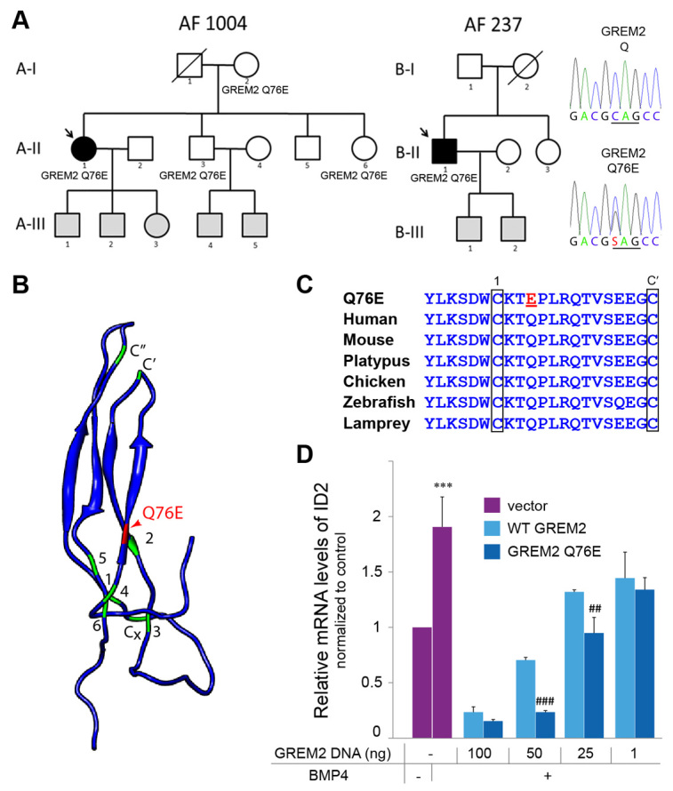
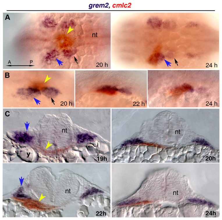
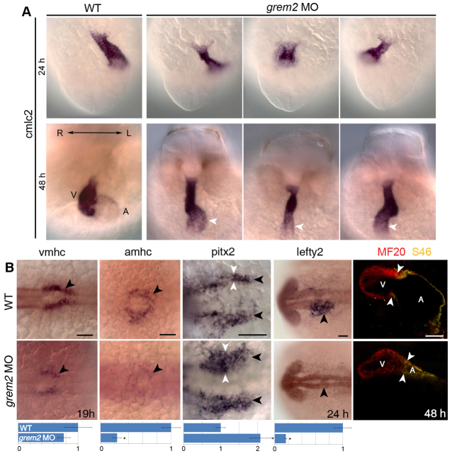
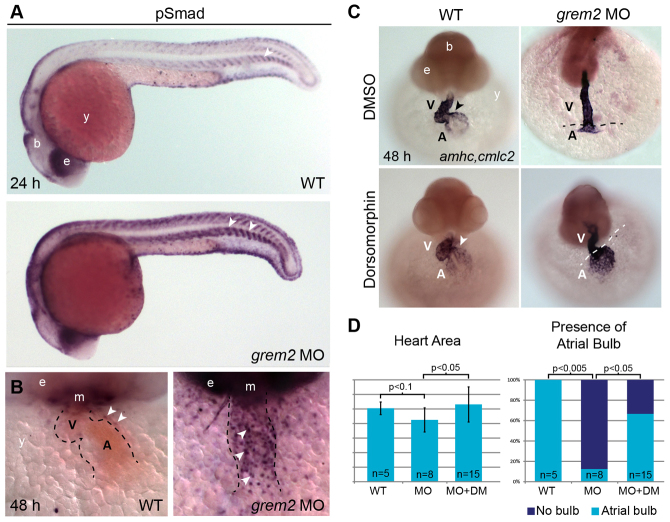
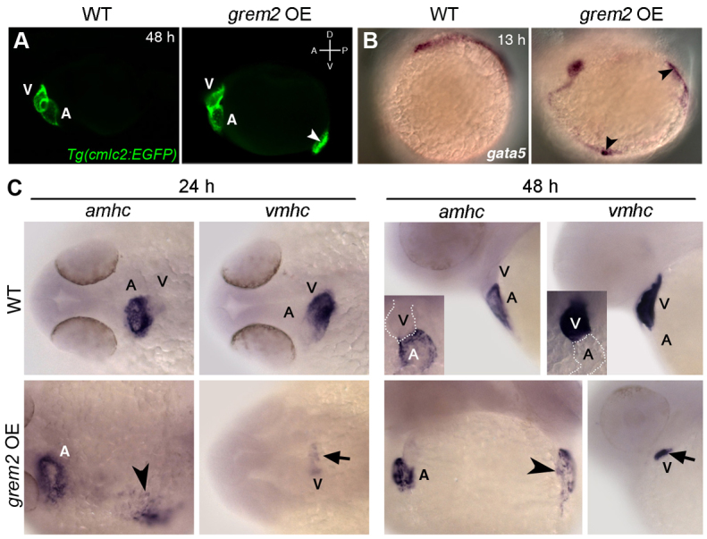
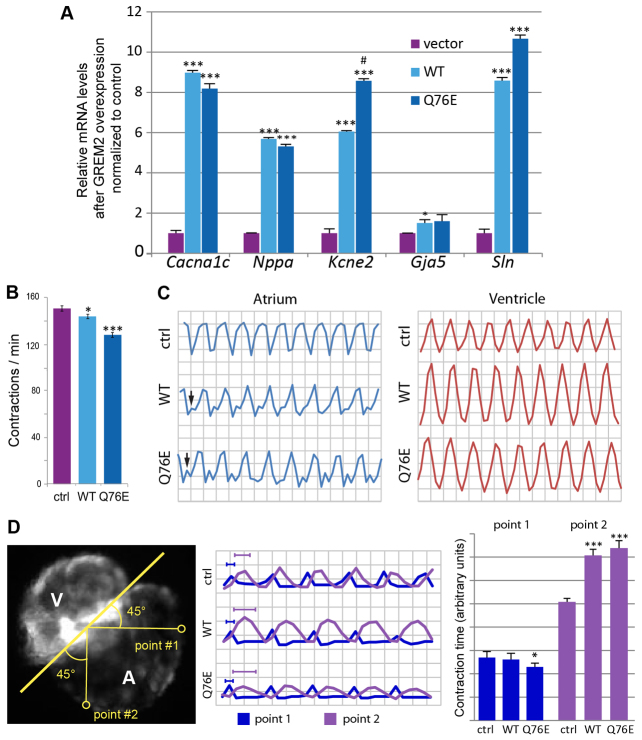
Atrial fibrillation (AF) is the most common cardiac arrhythmia and carries a significant risk of stroke and heart failure. The molecular etiologies of AF are poorly understood, leaving patients with limited therapeutic options. AF has been recognized as an inherited disease in almost 30% of patient cases. However, few genetic loci have been identified and the mechanisms linking genetic variants to AF susceptibility remain unclear. By sequencing 193 probands with lone AF, we identified a Q76E variant within the coding sequence of the bone morphogenetic protein (BMP) antagonist gremlin-2 (GREM2) that increases its inhibitory activity. Functional modeling in zebrafish revealed that, through regulation of BMP signaling, GREM2 is required for cardiac laterality and atrial differentiation during embryonic development. GREM2 overactivity results in slower cardiac contraction rates in zebrafish, and induction of previously identified AF candidate genes encoding connexin-40, sarcolipin and atrial natriuretic peptide in differentiated mouse embryonic stem cells. By live heart imaging in zebrafish overexpressing wild-type or variant GREM2, we found abnormal contraction velocity specifically in atrial cardiomyocytes. These results implicate, for the first time, regulators of BMP signaling in human AF, providing mechanistic insights into the pathogenesis of the disease and identifying potential new therapeutic targets.


Figures

Fig. 1.
Fig. 1.
Identification of GREM2 variant in probands with lone AF. (A) Pedigrees of two families with AF and probands heterozygous for the GREM2 c.226C>G variant allele that segregates in one of the families. Symbols indicate phenotypes as follows: solid, positive for AF; open, unaffected at >65 years of age. Due to the variable expressivity of AF and late onset of symptoms, patients age <65 years are considered to be of undetermined phenotype and are indicated by gray shading (A-III, B-III). Chromatographs are of proband carrying the Q76E mutation in GREM2 and wild-type control. (B) Protein model of GREM2 indicates the position of the amino acid substitution (red). The location of the six cysteines of the cystine knot are marked in green. Two additional cysteines C' and C” linking adjacent loops are indicated, as well as the unpaired Cx in the heel of the cysteine knot. The Q76E substitution is immediately adjacent to the conserved first cysteine (C73) of the cystine-knot motif at the base of the first of the two loops. (C) Sequence alignment shows that the Q76E substitution is in a highly conserved glutamine (Q) residue across species. (D) Relative expression levels of ID2 in HEK293 cells transfected with decreasing amounts of plasmids expressing wild-type GREM2 or GREM2-Q76E and treated with BMP4 recombinant protein. GREM2-Q76E decreased ID2 expression at lower levels than wild-type GREM2, suggesting that the variant was more potent at antagonizing BMP than wild-type GREM2. ***P<0.0001 versus no BMP4; ##P<0.01, ###P<0.0001 versus wild-type GREM2.
Fig. 2.
Fig. 2.
Expression ofgrem2in pharyngeal arch primordia during cardiac tube migration. (A,B) Whole mount images of grem2 (purple)- and cmlc2 (red)-stained embryos in dorsal (A) and lateral (B) views, anterior to the left. grem2 is expressed in the first two pharyngeal arches (blue arrows point to the left first arch, black arrows to the left second arch) as the cardiac progenitors begin to assemble in the midline. During cardiac jogging to the left, the heart tube (yellow arrowhead) is positioned next to grem2-expressing pharyngeal mesoderm and subsequently passes ventrally to the grem2-expression domain in the left first pharyngeal arch. (C) Cross-sections of zebrafish embryos double-labeled for grem2 and cmlc2. h, hours post fertilization; A, anterior; P, posterior; nt, neural tube; y, yolk.
Fig. 3.
Fig. 3.
Loss of Grem2 leads to cardiac jogging and looping defects, aberrant expression ofpitx2and abnormal development of cardiac chambers. (A) In situ hybridization analysis using cmlc2 riboprobe shows that in wild-type (WT) embryos the heart jogs leftward, whereas cardiac morphogenesis is randomized in grem2 morphants (grem2 MO). The cardiac tube, consisting of a single atrium (A, white arrowheads) and a single ventricle (V), is thinner, shorter and fails to loop in grem2 morphants. Left/Right (L/R) axis orientation is indicated. (B) In situ hybridization analysis using vmhc and amhc riboprobes shows that loss of Grem2 leads to lower expression levels of both vmhc and amhc, with amhc being essentially absent at 19 hpf (arrowheads). Pitx2 expression (black arrowheads) at 19 hpf is enhanced (white arrowheads), whereas lefty2 expression at 24 hpf is abolished (black arrowheads). Quantification of expression levels in wild-type and morphant embryos is shown below the corresponding images (expression is relative to wild type, which was set as arbitrary value 1). Immunofluorescence analysis using MF20 (red, labels both ventricular and atrial cardiomyocytes) and S46 (green, labels only atrial cardiomyocytes, which appear yellow) antibodies shows abnormal development of both chambers, with the atrium being more deformed and reduced in size than the ventricle. Arrowheads mark the position of the atrio-ventricular boundary. Scale bars: 50 μm. *P<0.05.
Fig. 4.
Fig. 4.
Loss of Grem2 leads to increased BMP signaling and cardiac defects that can be reversed by the BMP inhibitor dorsomorphin. (A) Whole mount immunohistochemistry using antibodies recognizing the phosphorylated forms of Smads1/5/8 shows stronger pSmad protein staining in grem2 morphants than in controls. Arrowhead marks somite boundaries. (B) Frontal close up views of the heart after pSmad1/5/8 antibody staining at 48 hpf shows sharply increased nuclear staining in cardiomyocytes of Grem2-depleted embryos. The heart is outlined by dotted lines; arrowheads mark pSmad-positive nuclei. (C) Wild-type (WT) embryos and grem2 morphants were incubated with dorsomorphin or its vehicle DMSO between 16 and 48 hpf and stained at 48 hpf with cmlc2 and amhc probes to visualize the ventricle (V) and atrium (A). Dorsomorphin treatment restored atrial patterning and differentiation (arrowheads point to the atrioventricular boundary in wild-type hearts; dotted lines demarcate ventricle-atrium boundary in grem2 morphants). (D) Quantification of heart size (Heart Area) and atrial bulb restoration (Presence of Atrial Bulb) in DMSO-treated controls (WT), DMSO-treated grem2 morphants (MO) and grem2 morphants treated with dorsomorphin (MO+DM). Dorsomorphin treatment restores atrial development. Error bars represent s.d. P-values and number of embryos analyzed (n) are indicated. b, brain; e, eye; m, mouth; y, yolk.
Fig. 5.
Fig. 5.
Overexpression of Grem2 induces ectopic atrial myocardium. (A) Lateral view of live wild-type (WT) and grem2 mRNA-injected (15 pg) cmlc2-egfp transgenic embryos (grem2 OE) at 48 hpf shows ectopic cardiac tissue (arrowhead). Anterior-posterior (A/P) and dorsal-ventral (D/V) axes are indicated. (B) In situ hybridization shows ectopic sites expressing gata-5 along the paraxial mesoderm (arrowheads). (C) WT zebrafish embryos were injected with grem2 mRNA at the 1-4 cell stage; non-injected embryos served as controls. At 24 hpf, Grem2 overexpression (OE) led to enlarged or ectopic (arrowhead) amhc expression in 100% of injected embryos, whereas vmhc expression was reduced (arrow) or absent in 54.4%. These results remained consistent at 48 hpf, with 96% of embryos displaying ectopic amhc expression (arrowhead), and 52.2% displaying reduced vmhc expression (arrow). Insets display enlarged frontal images. Normal atrium (A) expressing amhc and ventricle (V) expressing vmhc are marked; dorsal (24 hpf) and lateral (48 hpf) views; anterior is to the left in all panels.
Fig. 6.
Fig. 6.
GREM2 activity regulates cardiac rhythm. (A) Stable mouse embryonic stem cell lines, generated with empty vector (control) or vectors expressing wild-type GREM2 or GREM2-Q76E, were allowed to form embryoid bodies. RNA was then isolated at differentiation day 8 and analyzed by qRT-PCR. The results showed that overexpression of GREM2 and GREM2-Q76E leads to upregulation of genes encoding calcium channel, voltage-dependent, L type, alpha 1C subunit (Cacna1c), natriuretic atrial peptide (Nppa), potassium voltage-gated channel subfamily E member 2 (Kcne2), connexin-40 (Gja5) and sarcolipin (Sln). (B) Quantification of cardiac contraction rates in vivo in Tg(cmlc2:EGFP) zebrafish embryos overexpressing human GREM2 and GREM2-Q76E variant. Both wild-type GREM2 and GREM2-Q76E slowed contraction rates, but the effect was more pronounced in GREM2-Q76E-injected embryos. (C) Digital cardiography of Tg(cmlc2:EGFP) zebrafish at 48 hpf showed regular ventricular rhythm and discordant atrial rhythm in wild-type GREM2 and variant GREM2-Q76E injected embryos (arrows). (D) Tracking of two points in the proximal (1) and distal (2) atrium over time with quantification of time spent by each point in systole showed that the time spent in systole was not disrupted in the proximal point, but was significantly increased in the distal point in both wild-type (WT) and variant-injected embryos, suggesting a possible deterioration of the contraction wave as it traveled across the atrium. *P<0.05, **P<0.001, ***P<0.0001 versus control; #P<0.01, ##P<0.001 versus wild-type GREM2.

Similar articles

Cited by

References

    1. Abraham R. L., Yang T., Blair M., Roden D. M., Darbar D. (2010). Augmented potassium current is a shared phenotype for two genetic defects associated with familial atrial fibrillation. J. Mol. Cell. Cardiol. 48, 181-190 - PMC - PubMed
    1. Antzelevitch C., Pollevick G. D., Cordeiro J. M., Casis O., Sanguinetti M. C., Aizawa Y., Guerchicoff A., Pfeiffer R., Oliva A., Wollnik B., et al. (2007). Loss-of-function mutations in the cardiac calcium channel underlie a new clinical entity characterized by ST-segment elevation, short QT intervals, and sudden cardiac death. Circulation 115, 442-449 - PMC - PubMed
    1. Avsian-Kretchmer O., Hsueh A. J. (2004). Comparative genomic analysis of the eight-membered ring cystine knot-containing bone morphogenetic protein antagonists. Mol. Endocrinol. 18, 1-12 - PubMed
    1. Beck H., Semisch M., Culmsee C., Plesnila N., Hatzopoulos A. K. (2008). Egr-1 regulates expression of the glial scar component phosphacan in astrocytes after experimental stroke. Am. J. Pathol. 173, 77-92 - PMC - PubMed
    1. Bisgrove B. W., Essner J. J., Yost H. J. (1999). Regulation of midline development by antagonism of lefty and nodal signaling. Development 126, 3253-3262 - PubMed

Publication types

MeSH terms