

Review
doi: 10.1007/82_2012_289.
Cell signaling pathways in vertebrate lens regeneration
Affiliations
- PMID: 23224710
- PMCID: PMC4304700
- DOI: 10.1007/82_2012_289
Item in Clipboard
Review
Cell signaling pathways in vertebrate lens regeneration
Curr Top Microbiol Immunol.
2013.
Abstract
Certain vertebrates are capable of regenerating parts of the eye, including the lens. Depending on the species, two principal forms of in vivo lens regeneration have been described wherein the new lens arises from either the pigmented epithelium of the dorsal iris or the cornea epithelium. These forms of lens regeneration are triggered by retinal factors present in the eye. Studies have begun to illuminate the nature of the signals that support lens regeneration. This review describes evidence for the involvement of specific signaling pathways in lens regeneration, including the FGF, retinoic acid, TGF-beta, Wnt, and Hedgehog pathways.
Figures
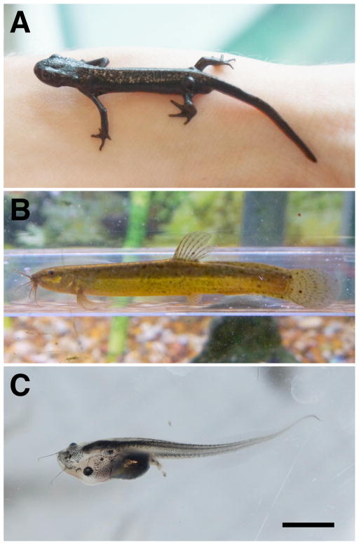
Examples of various animals capable of regenerating the lens of the eye. A). The newt Cynops pyrrhogaster (the Japanese Fire Belly Newt). B). The cobitid fish, Misgurnus anguillicaudatus (the Japanese Weather Loach) C). The tadpole larvae of the frog Xenopus laevis (the South African Clawed Frog). See text for further details. Scale bar equals 7mm for A, 15mm for B and 6mm for C.
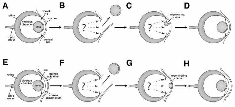
Diagrams illustrating the two main processes of lens regeneration. A–D). Steps involved in Wolffian lens regeneration that take place in certain newts and salamanders. This process takes place within the dorsal iris of the adult eye following removal of the lens (B) (i.e., the new lens is derived from pigmented iris epithelial cells that undergo transdifferentiation). Dashed arrows in B and C indicate key retinal signals that support the process of lens regeneration. E–H). Steps involved in cornea lens regeneration that takes place in frogs of the genus Xenopus and the salamander Hynobius unnangso. The process takes place within cells of the basal layer of the larval cornea epithelium following removal of the lens and perforation of the cornea endothelium (F). Dashed arrows in F and G indicate key retinal signals that support the process of lens regeneration.
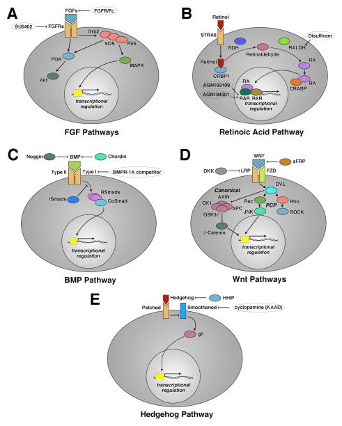
Highly simplified summary diagrams of various signaling pathways known to play roles during vertebrate lens regeneration. Various pathways and pathway members are as labeled. Not all known elements are included here for the sake of simplicity. Specific activators and inhibitors used to manipulate these pathways are also included, and specific synthetic agents are enclosed in ovals. See text for further details.
Similar articles
-
Lens regeneration from the cornea requires suppression of Wnt/β-catenin signaling.Exp Eye Res. 2016 Apr;145:206-215. doi: 10.1016/j.exer.2016.01.003. Epub 2016 Jan 8. Exp Eye Res. 2016. PMID: 26778749 Free PMC article.
-
Retinoic acid regulation by CYP26 in vertebrate lens regeneration.Dev Biol. 2014 Feb 15;386(2):291-301. doi: 10.1016/j.ydbio.2013.12.036. Epub 2013 Dec 30. Dev Biol. 2014. PMID: 24384390 Free PMC article.
-
The cellular and molecular bases of vertebrate lens regeneration.Int Rev Cytol. 2003;228:195-265. doi: 10.1016/s0074-7696(03)28005-0. Int Rev Cytol. 2003. PMID: 14667045 Review.
-
Conservation of fibroblast growth factor function in lens regeneration.Proc Natl Acad Sci U S A. 1997 Dec 9;94(25):13701-6. doi: 10.1073/pnas.94.25.13701. Proc Natl Acad Sci U S A. 1997. PMID: 9391089 Free PMC article.
-
[Cell sources, regulatory factors and gene expression in the regeneration of the crystalline lens and retina in vertebrate animals].Izv Akad Nauk Ser Biol. 1996 May-Jun;(3):298-318. Izv Akad Nauk Ser Biol. 1996. PMID: 8755029 Review. Russian.
Cited by
-
Lens regeneration from the cornea requires suppression of Wnt/β-catenin signaling.Exp Eye Res. 2016 Apr;145:206-215. doi: 10.1016/j.exer.2016.01.003. Epub 2016 Jan 8. Exp Eye Res. 2016. PMID: 26778749 Free PMC article.
-
Lens regeneration: scientific discoveries and clinical possibilities.Mol Biol Rep. 2021 May;48(5):4911-4923. doi: 10.1007/s11033-021-06489-5. Epub 2021 Jun 18. Mol Biol Rep. 2021. PMID: 34143397 Review.
-
Lens regeneration in humans: using regenerative potential for tissue repairing.Ann Transl Med. 2020 Nov;8(22):1544. doi: 10.21037/atm-2019-rcs-03. Ann Transl Med. 2020. PMID: 33313289 Free PMC article. Review.
-
Diverse Evolutionary Origins and Mechanisms of Lens Regeneration.Mol Biol Evol. 2018 Jul 1;35(7):1563-1575. doi: 10.1093/molbev/msy045. Mol Biol Evol. 2018. PMID: 29579253 Free PMC article. Review.
-
Go ahead, grow a head! A planarian's guide to anterior regeneration.Regeneration (Oxf). 2016 Jun 24;3(3):139-55. doi: 10.1002/reg2.56. eCollection 2016 Jun. Regeneration (Oxf). 2016. PMID: 27606065 Free PMC article. Review.
References
-
- Ang SJ, Stump RJ, Lovicu FJ, McAvoy JW. Spatial and temporal expression of Wnt and Dickkopf genes during murine lens development. Gene Expr Patterns. 2004;4:289–295. - PubMed
-
- Arresta E, Bernardini S, Gargioli C, Filoni S, Cannata SM. Lens-forming competence in the epidermis of Xenopus laevis during development. J Exp Zool. 2005;303A:1–12. - PubMed
-
- Blobe G, Liu X, Fang SJ, How T, Lodish HF. A novel mechanism for regulating transforming growth factor β (TGF-β) signaling. Functional modulation of type III TGF-b receptor expression through interaction with the PDZ domain protein. GIPC J Biol Chem. 2001;276:39608–39617. - PubMed
Publication types
MeSH terms
Substances
Grants and funding
LinkOut - more resources
Full Text Sources
Other Literature Sources
