

The shape of the proximal femur influences acetabular wear patterns over time
- PMID: 23224910
- PMCID: PMC3549172
- DOI: 10.1007/s11999-012-2720-x
The shape of the proximal femur influences acetabular wear patterns over time
Abstract
Background: Femoroacetabular impingement has been proposed as a cause of early osteoarthritis, but it is not known how this develops over time or whether the shape of the proximal femur influences this risk.
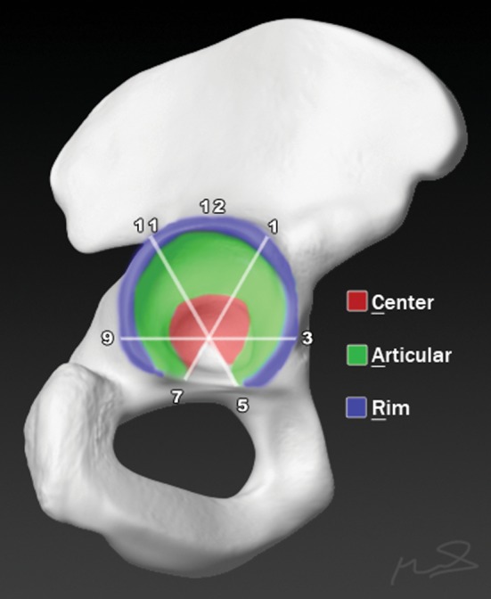
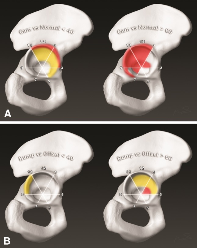
Questions/purposes: (1) Which areas of the acetabulum are worn more frequently by individuals with a cam deformity of the proximal femur? (2) Do observed acetabular wear patterns differ based on the etiology of the cam deformity? (3) Do wear patterns of individuals with a cam deformity differ based on an individual's age?
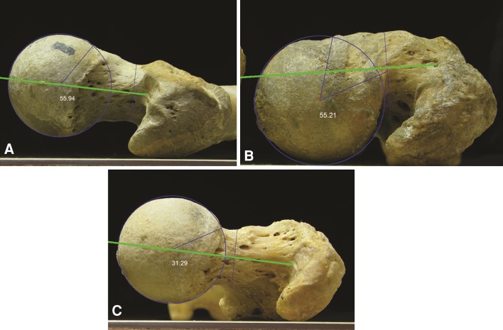
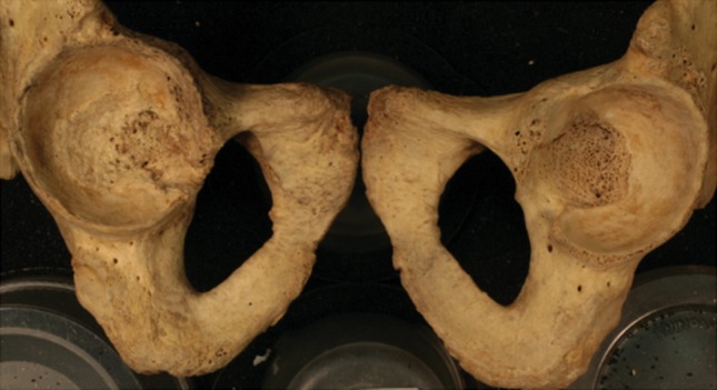
Methods: We examined 645 corresponding femora and acetabuli from the Hamann-Todd Osteological Collection and determined the offset and alpha angle using photographs; 370 specimens met inclusion criteria and were examined for signs of wear and the locations of wear were recorded. Specimens were separated into eight subgroups based on age either younger than 40 years or older than 60 years, alpha angle greater or less than 55°, and degree of anterior head-neck offset. We compared the prevalence of wear between groups in each location.
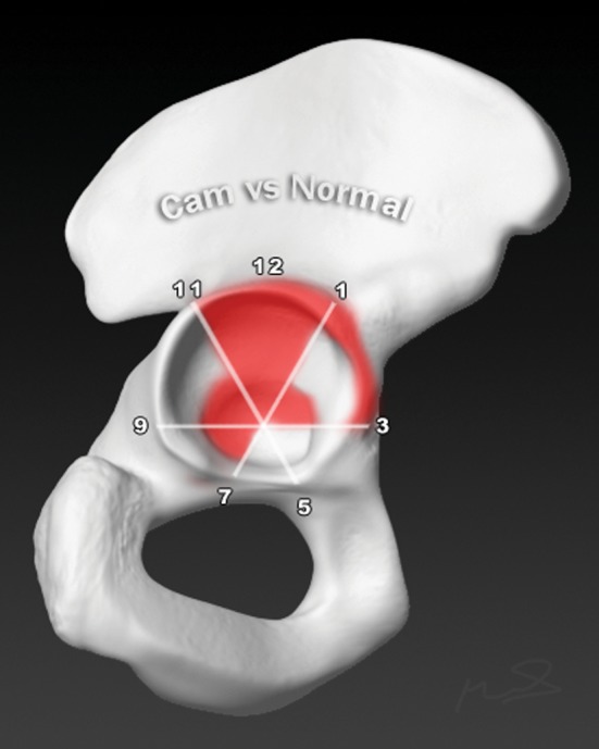
Results: Individuals with abnormal geometry of the proximal femur demonstrated different wear patterns from individuals with normal geometry. There were few differences in wear patterns identified based on the etiology of the femoral deformity. Abnormal femoral geometry was associated with more frequent wear primarily at the anterosuperior acetabulum for individuals younger than 40 years of age and globally for individuals older than 60 years of age.
Conclusion: Femoral geometry appears to influence the pattern of acetabular wear occurring over time.
Figures
Similar articles
-
The Pattern of Acetabular Cartilage Wear Is Hip Morphology-dependent and Patient Demographic-dependent.Clin Orthop Relat Res. 2019 May;477(5):1021-1033. doi: 10.1097/CORR.0000000000000649. Clin Orthop Relat Res. 2019. PMID: 30998630 Free PMC article.
-
Twelve percent of hips with a primary cam deformity exhibit a slip-like morphology resembling sequelae of slipped capital femoral epiphysis.Clin Orthop Relat Res. 2015 Apr;473(4):1212-23. doi: 10.1007/s11999-014-4068-x. Clin Orthop Relat Res. 2015. PMID: 25448326 Free PMC article.
-
Symptomatic femoroacetabular impingement: does the offset decrease correlate with cartilage damage? A pilot study.Clin Orthop Relat Res. 2013 Jul;471(7):2173-82. doi: 10.1007/s11999-013-2812-2. Clin Orthop Relat Res. 2013. PMID: 23361934 Free PMC article.
-
[Femoroacetabular impingement: a new direction in the diagnosis and treatment of the hip joint].Harefuah. 2011 Feb;150(2):148-52, 205. Harefuah. 2011. PMID: 22164944 Review. Hebrew.
-
More than just a bump: cam-type femoroacetabular impingement and the evolution of the femoral neck.Hip Int. 2011 Jan-Mar;21(1):1-8. doi: 10.5301/hip.2011.6288. Hip Int. 2011. PMID: 21279972 Review.
Cited by
-
The Pattern of Acetabular Cartilage Wear Is Hip Morphology-dependent and Patient Demographic-dependent.Clin Orthop Relat Res. 2019 May;477(5):1021-1033. doi: 10.1097/CORR.0000000000000649. Clin Orthop Relat Res. 2019. PMID: 30998630 Free PMC article.
-
The natural history of alpha angle in the last seventeen centuries.Arch Orthop Trauma Surg. 2022 Oct;142(10):2819-2825. doi: 10.1007/s00402-021-04268-2. Epub 2021 Nov 26. Arch Orthop Trauma Surg. 2022. PMID: 34825963
-
Femurs in patients with hip dysplasia have fundamental shape differences compared with cam femoroacetabular impingement.J Hip Preserv Surg. 2024 Feb 5;11(2):132-139. doi: 10.1093/jhps/hnae004. eCollection 2024 Jul. J Hip Preserv Surg. 2024. PMID: 39070210 Free PMC article.
References
-
- Cooperman DR, Wallensten R, Stulberg SD. Acetabular dysplasia in the adult. Clin Orthop Relat Res. 1983;175:79–85. - PubMed
-
- Ganz R, Parvizi J, Beck M, Leunig M, Nötzli H, Siebenrock KA. Femoroacetabular impingement: a cause for osteoarthritis of the hip. Clin Orthop Relat Res. 2003;417:112–120. - PubMed
MeSH terms
LinkOut - more resources
Full Text Sources
Other Literature Sources
Miscellaneous
