. 2013 Apr;30(4):541-52.
doi: 10.1007/s10585-012-9558-1. Epub 2012 Dec 6.

Integrin α3β1 regulates tumor cell responses to stromal cells and can function to suppress prostate cancer metastatic colonization

Affiliations

Integrin α3β1 regulates tumor cell responses to stromal cells and can function to suppress prostate cancer metastatic colonization

Afshin Varzavand et al. Clin Exp Metastasis. 2013 Apr.

Abstract

Integrin α3β1 promotes tumor cell adhesion, migration, and invasion on laminin isoforms, and several clinical studies have indicated a correlation between increased tumoral α3β1 integrin expression and tumor progression, metastasis, and poor patient outcomes. However, several other clinical and experimental studies have suggested that α3β1 can possess anti-metastatic activity in certain settings. To help define the range of α3β1 functions in tumor cells in vivo, we used RNAi to silence the α3 integrin subunit in an aggressive, in vivo-passaged subline of PC-3 prostate carcinoma cells. Loss of α3 integrin impaired adhesion and proliferation on the α3β1 integrin ligand, laminin-332 in vitro. Despite these deficits in vitro, the α3-silenced cells were significantly more aggressive in a lung colonization model in vivo, with a substantially increased rate of tumor growth that significantly reduced survival. In contrast, silencing the related α6 integrin subunit delayed metastatic growth in vivo. The increased colonization of α3-silenced tumor cells in vivo was recapitulated in 3D collagen co-cultures with lung fibroblasts or pre-osteoblast-like cells, where α3-silenced cells showed dramatically enhanced growth. The increased response of α3-silenced tumor cells to stromal cells in co-culture could be reproduced by fibroblast conditioned medium, which contains one or more heparin-binding factors that selectively favor the growth of α3-silenced cells. Our new data suggest a scenario in which α3β1 regulates tumor-host interactions within the metastatic tumor microenvironment to limit growth, providing some of the first direct evidence that specific loss of α3 function in tumor cells can have pro-metastatic consequences in vivo.


Figures
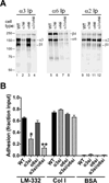
Fig. 1
Fig. 1
Specific silencing of α3 and α6 integrin subunits in GS698.Li prostate carcinoma cells. a Parental (WT), α3-silenced (α3si), α6-silenced (α6si), and doubly silenced cells (α3/α6si) were surface-labeled with biotin and extracted with 1% Triton X-100. Integrins α3β1, α6β4, or α2β1 were immunoprecipitated (Ip) from normalized lysates, and then analyzed by blotting with IRdye 800-streptavidin. b WT, α3si, α6si, and α3/α6si cells were allowed to adhere to wells coated with laminin-332 (LM-332), collagen I (Col I), or BSA for 20 min in serum-free medium. Non-adherent cells were removed by rinsing, and adherent cells were fixed and quantified by staining with crystal violet. Results are presented as a fraction of total cells input, as measured in poly-L-lysine control wells. Two independent trials that gave similar results were pooled (for total of 8 wells per cell type/condition) Error bars indicate S.E.M. *Significantly less than WT parental cells, ANOVA with Tukey-Kramer t test, (p<0.001); **Significantly less than WT (p<0.001) and significantly less than α3si (p<0.05), ANOVA with Tukey-Kramer t test.
Fig. 2
Fig. 2
Divergent functions for α3 and α6 integrins in metastatic colonization of GS689.Li prostate carcinoma cells. a Bioluminescence imaging (BLI) was used to visualize WT, α3si, α6si, and α3/α6si GS689.Li cells immediately after tail vein inoculation (Day 0) or 35 days later. Eight mice per cell type were injected, four of each group are depicted. Note that the photon flux color scale is 4 fold higher on week 5 than on day 0. b The average apparent tumor burden for each group was measured each week by BLI, as described in Materials and Methods. Statistical analysis: significantly different from WT parental cells, ANOVA with Dunnett t test (*p<0.05, **p<0.01 or lower). c Kaplan-Meier analysis of the percent survival to endpoint for the 8 mice per group (using criteria described in Materials and Methods) revealed significantly reduced survival times for α3si and α3/α6si cells (p<0.0002) and significantly increased survival time for α6si cells (p<0.003) as compared to WT parental cells. d Kaplan-Meier analysis of extra-thoracic colonization revealed significantly reduced colonization for α6si cells compared to the other groups (p<0.004).
Fig. 3
Fig. 3
Severely impaired spreading of α3 silenced cells on LM-332. a & b WT and α3si cells were plated in serum-free medium on LM-332-coated glass bottom culture dishes. After 30 min for cell attachment and spreading, cells were photographed with a 20X objective. c 50 cells of each type were quantified using ImageJ to measure cell area. The α3si cells were significantly less well spread, p < 0.0001, unpaired t test.
Fig. 4
Fig. 4
Analysis of in vitro GS689.Li tumor cell proliferation. WT and α3si cells were plated in serum-free medium in 96 well plates in (a) uncoated wells, (b) laminin-332-coated wells, or (c) collagen I-coated wells. Replicate plates were analyzed on successive days by a WST colorimetric assay (6 wells/cell type per time point). Statistical analysis: *value for WT cells was significantly higher than that of α3si cells (p<0.001, unpaired t test).
Fig. 5
Fig. 5
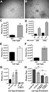
α3 integrin negatively regulates tumor cell growth in 3D co-cultures with stromal cells. a & b WT or α3si GS689.Li cells (3,000 cells per well) were plated on 3D collagen in which 10,000 MRC-5 human lung fibroblasts per well had been embedded. After 17 d, cells were photographed with a 4X objective. c A co-culture experiment was performed as described in a & b, and tumor cell number was quantified using bioluminescence imaging (BLI) after 28 d. *Significantly greater than WT, p<0.0001, unpaired t test, (n=5 wells/cell type). d WT or α3si tumor cells (3,000/well) were plated on 3D collagen in which 3,000 or 10,000 MRC-5 human lung fibroblasts per well had been embedded. After 24 d, tumor cell number was quantified by BLI. *Significantly greater than WT, p<0.0001, unpaired t test (n=6 wells/cell type). e A co-culture experiment was performed as described in a & b, but comparing WT to α3si-2 cells, in which residual cell surface α3 is ~40% of WT α3 expression. After 28 d, tumor cell number was quantified by BLI. *Significantly greater than WT, p<0.001, unpaired t test, (n=10 wells/cell type). f A co-culture experiment was performed as in a & b, except that 10,000 MC3T3-E1 pre-osteoblast-like cells per well were embedded in collagen instead of 10,000 MRC-5 fibroblasts. After 18 d, tumor cell number was quantified by BLI. *Significantly greater than WT, p<0.0001, unpaired t test (n=6 wells/cell type). g A co-culture experiment was performed as in a & b, but comparing untreated WT tumor cells (WT), WT tumor cells treated with 10 µg/ml A3-IIF5 anti-α3 integrin antibody (WT anti-α3), and α3si tumor cells (α3si). In addition, MRC-5 cells were omitted from one set each of WT and α3si wells (no MRC-5). After 16 d, tumor cell number was quantified by BLI. Statistical analysis: significantly greater than untreated WT wells, *p<0.05, **p<0.01, ANOVA with Tukey-Kramer t test (n=6 wells/cell type). h LNCaP C4-2B prostate carcinoma cells transduced with empty vector (C4-2B VEC) or with an α3 integrin expression vector (C4-2B α3) were plated at 3,000 cells per well on (i) 3D collagen in which 10,000 MRC-5 fibroblasts per well had been embedded, or (ii) on 3D collagen lacking MRC-5 cells (no MRC-5). After 26 d, tumor cell number was quantified by BLI. Statistical analysis: the indicated comparisons were significant at *p<0.01, **p<0.001, ANOVA with Tukey-Kramer t test (n=6 wells/cell type); Graphs in C-H all depict mean ± S.E.M.
Fig. 6
Fig. 6
MRC-5 fibroblasts secrete one or more heparin-binding factors that selectively promote the growth of α3-silenced GS689.Li tumor cells. Freshly confluent cultures of MRC-5 fibroblasts were used to condition serum-free medium for 3 days, and then the medium was harvested and used in tumor cell growth assays. a WT and α3si GS689.Li tumor cells (3,000 cells/well) were plated on 3D collagen in varying dilutions of MRC-5 conditioned medium ranging from 0% (non-conditioned medium) to 33% (MRC-5 conditioned medium diluted 1:3 in non-conditioned medium). Wells were refed after 1 wk with fresh dilutions of MRC-5 conditioned medium. After 2 wks, tumor cell number was quantified by bioluminescence imaging (BLI). The α3si cells displayed a dramatically enhanced dose-response to MRC-5 conditioned medium. *Significantly greater than WT wells, p<0.0001, unpaired t test (n=4 wells/cell type). Values plotted are mean ± S.E.M. b MRC-5 conditioned medium was fractionated on heparin sepharose, as described in Materials and Methods. Fractions were diluted 1:8 and then used in growth assays of WT and α3si tumor cells on 3D collagen as in panel a. SM, starting material; FT, flow through; 0.15 M – 2.0M, NaCl elution steps of the heparin sepharose column, SFM, non-conditioned, serum-free medium. Values plotted are mean ± S.E.M. for 3 wells/cell type/condition.

Similar articles

Cited by

References

    1. Siegel R, Naishadham D, Jemal A. Cancer statistics, 2012. CA Cancer J Clin. 2012;62:10–29. - PubMed
    1. Goel HL, Alam N, Johnson INS, Languino LR. Integrin signaling aberrations in prostate cancer. Am J Transl Res. 2009;1:211–220. - PMC - PubMed
    1. Goel HL, Li J, Kogan S, Languino LR. Integrins in prostate cancer progression. Endocr Relat Cancer. 2008;15:657–664. - PMC - PubMed
    1. Knudsen BS, Miranti CK. The impact of cell adhesion changes on proliferation and survival during prostate cancer development and progression. J Cell Biochem. 2006;99:345–361. - PubMed
    1. Sroka IC, Anderson TA, McDaniel KM, Nagle RB, Gretzer MB, et al. The laminin binding integrin alpha6beta1 in prostate cancer perineural invasion. J Cell Physiol. 2010;224:283–288. - PMC - PubMed

Publication types

MeSH terms

Substances