Review
. 2013:768:197-211.
doi: 10.1007/978-1-4614-5107-5_12.

Relationship of GW/P-bodies with stress granules

Affiliations
Review

Relationship of GW/P-bodies with stress granules

Georg Stoecklin et al. Adv Exp Med Biol. 2013.

Abstract

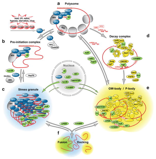
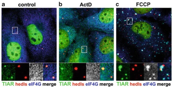
Whereas P-bodies are intimately linked to the cytoplasmic RNA decay machinery, stress granules harbor stalled translation initiation complexes that accumulate upon stress-induced translation arrest. In this Chapter, we reflect on the relationship between P-bodies and stress granules. In mammalian cells, the two structures can be clearly distinguished from each other using specific protein or RNA markers, but they also share many proteins and mRNAs. While the formation of P-bodies and stress granules is coordinately triggered by stress, their assembly appears to be regulated independently by different pathways. Under certain types of stress, P-bodies frequently dock with stress granules, and overexpressing certain proteins that localize to both structures can cause P-body/stress granule fusion. Currently available data suggest that these self-assembling compartments are controlled by flux of mRNAs within the cytoplasm, and that their assembly mirrors the translation and degradation rates of their component mRNAs.


Figures

Fig. 12.1
Fig. 12.1
Scheme of mRNP complexes forming stress granules and P-bodies. (a) Actively translating mRNAs are capped, polyadenylated and form polysomes. (b) Under conditions of severe stress, global mRNA translation is inhibited by phosphorylation of eIF2 through activation of stress-responsive kinase PERK, GCN2, HRI or PKR. As a consequence, polysomes disassemble, and translation pre-initiation complexes accumulate. The protein phosphatase PP1 together with its regulatory subunit Gadd34 dephosphorylates eIF2 and thereby re-activates translation. (c) RNA-binding proteins such as TIA1 and G3BP contain aggregation-prone domains that drive the assembly of stalled pre-initiation complexes into cytoplasmic stress granules. The conjugation of N-acetylglucosamine (GlcNAc) to ribosomal proteins is also important for stress granule assembly. When stressful conditions are overcome, the chaperone Hsp70 facilitates disassembly of stress granules. (d) mRNAs are specifically degraded if they have acquired a premature termination codon (PTC), associate with miRNAs or contain AU-rich elements (AREs). Such mRNAs are either cleaved by an endonculease or subject to rapid deadenylation, which induces decapping and degradation through the 5′-3′ exoribonuclease Xrn1. (e) mRNAs targeted for decay together with key enzymes of cytoplasmic RNA degradation assemble in processing (P)-bodies. The RNA helicase Rck and aggregation-prone proteins such as Pat1, Edc3 and Lsm4 are important for P-body formation. miRNAs can also suppress the translation of target mRNAs by recruiting them to P-bodies, from where such mRNAs can also exit and re-engage in translation. (f) P-bodies are frequently observed in close physical contact to stress granules as if they were docking. The over-expression of certain RNA-binding proteins further enhances the association between P-bodies and stress granules, and causes fusion of the two structures. Such activity is observed for the cytoplasmic polyadenylation element binding protein (CPEB1) as well as for tristetraprolin (TTP) and butyrate response factor-1 (BRF1), two zinc finger proteins that accelerate the degradation of ARE-containing mRNAs. Stress granule proteins are shown in blue, P-body proteins in yellow, and proteins that localize to both structures are shown in green
Fig. 12.2
Fig. 12.2
Immunofluorescence micrograph of P-bodies docking to stress granules. African green monkey COS7 kidney cells were (a) grown under normal conditions, (b) exposed to 5 μg/mL actinomycin D (ActD) for 1 h, or (c) treated with carbonyl cyanide p-trifluoromethoxyphenylhydrazone (FCCP) in glucose-free media for 1 h. Cells were then fixed and stained for TIAR (green), the stress-granule specific marker protein eIF4G (blue), and the P-body-specific protein Hedls/GE-1 (red). Actinomycin D treatment does not induce stress granules but causes TIAR accumulation at P-bodies, whereas FCCP treatment triggers stress granule formation and promotes docking of P-bodies with stress granules

Similar articles

Cited by

References

    1. Aizer A, Brody Y, Ler LW, Sonenberg N, Singer RH, Shav-Tal Y. The dynamics of mammalian P body transport, assembly, and disassembly in vivo. Mol Biol Cell. 2008;19:4154–4166. - PMC - PubMed
    1. Andrei MA, Ingelfinger D, Heintzmann R, Achsel T, Rivera-Pomar R, Luhrmann R. A role for eIF4E and eIF4E-transporter in targeting mRNPs to mammalian processing bodies. RNA. 2005;11:717–727. - PMC - PubMed
    1. Arimoto K, Fukuda H, Imajoh-Ohmi S, Saito H, Takekawa M. Formation of stress granules inhibits apoptosis by suppressing stress-responsive MAPK pathways. Nat Cell Biol. 2008;10:1324–1332. - PubMed
    1. Athanasopoulos V, Barker A, Yu D, Tan AH, Srivastava M, Contreras N, Wang J, Lam KP, Brown SH, Goodnow CC, Dixon NE, Leedman PJ, Saint R, Vinuesa CG. The ROQUIN family of proteins localizes to stress granules via the ROQ domain and binds target mRNAs. FEBS J. 2010;277:2109–2127. - PubMed
    1. Baez MV, Boccaccio GL. Mammalian Smaug is a translational repressor that forms cytoplasmic foci similar to stress granules. J Biol Chem. 2005;280:43131–43140. - PubMed

Publication types

MeSH terms

LinkOut - more resources