. 2013 Mar;30(3):878-88.
doi: 10.1007/s11095-012-0929-8. Epub 2012 Dec 7.

(89)Zr-labeled paramagnetic octreotide-liposomes for PET-MR imaging of cancer

Affiliations

(89)Zr-labeled paramagnetic octreotide-liposomes for PET-MR imaging of cancer

Diane S Abou et al. Pharm Res. 2013 Mar.

Abstract

Purpose: Dual-modality PET/MR platforms add a new dimension to patient diagnosis with high resolution, functional, and anatomical imaging. The full potential of this emerging hybrid modality could be realized by using a corresponding dual-modality probe. Here, we report pegylated liposome (LP) formulations, housing a MR T(1) contrast agent (Gd) and the positron-emitting (89)Zr (half-life: 3.27 days), for simultaneous PET and MR tumor imaging capabilities.

Methods: (89)Zr oxophilicity was unexpectedly found advantageous for direct radiolabeling of preformed paramagnetic LPs. LPs were conjugated with octreotide to selectively target neuroendocrine tumors via human somatostatin receptor subtype 2 (SSTr2). (89)Zr-Gd-LPs and octreotide-conjugated homolog were physically, chemically and biologically characterized.

Results: (89)Zr-LPs showed reasonable stability over serum proteins and chelator challenges for proof-of-concept in vitro and in vivo investigations. Nuclear and paramagnetic tracking quantified superior SSTr2-recognition of octreotide-LP compared to controls.

Conclusions: This study demonstrated SSTr2-targeting specificity along with direct chelator-free (89)Zr-labeling of LPs and dual PET/MR imaging properties.


Figures

Fig. 1
Fig. 1
Characterization of OCT coupled to DSPE-PEG2000-CO2H (a and b) and to LPs (c and d). (a) C4-HPLC chromatogram of OCT and DSPE-PEG2000-CO2H coupling performed at pH 6 with EDC and S-NHS and acquired using electrospray ionization-QTOF-MS (ESI) and UV-280 nm detection. (b) Mass spectrum of mono-coupled DSPE-PEG2000-N-OCT, eluted at 31 min (chromatogram A). (c) OL conjugation scheme starting with Gd-CL; UC: ultracentrifugation. (d) HPLC evaluation of coupling efficiency by subtracting post-reaction supernatant (SN) OCT concentration from the initial OCT reaction concentration.
Fig. 2
Fig. 2
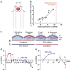
Direct Zr adsorption on lipid membranes. (a) Scheme of Zr binding to adjacent lipidic phosphate heads. (b) The surface tension Δπ increases upon addition of Zr (0.2 μmol) at various initial surface pressures (πi). (c) Scheme of DSPC monolayer membrane states (#1 to #4) characterized by Δπ variation. Δπ is: #1) positive and increasing when lipids adsorb substrates (Zr, red, or DFO, blue); #2) dropped when the membrane saturates in adsorbing substances; #3) stable when no interaction occurs with the membrane; and, #4) negative and decreasing when Zr-DFO complexes desorb from the membrane. (d and e) Δπ variation upon adsorption of Zr (red line) or DFO (blue line), followed by Zr-DFO desorption upon the addition of DFO or Zr.
Fig. 3
Fig. 3
Stability study of 89Zr-LPs. (a) Size exclusion chromatogram of the 48 h incubated mixture: mouse serum protein (MSP) and 89Zr-LPs. (b) LPs radioactivity stability profile after incubation and purification at 24; 48; 96 h with MSP or BSA (5%w/v); n≥2.
Fig. 4
Fig. 4
(a) 50 h p.i. T1-weighted MR scan of mice implanted with wild type (wt) (→) and SSTr2-postive tumors (◆→), injected with OL and CL (35–40 μCi; 18.5 mM–2.78 μmol Gd-DTPA-BiSA; 43.5 mM–6.53 μmol lipids). (b) Voxel-wise analysis of tumors normalized (to standard) intensity. Selective MR enhancement is achieved using targeted OL. (c) Percentage of contrast enhancement (prescan to 50 h p.i.) of tumor ROI normalized to water standard. Nearly two fold enhancement is observed using targeted OL (n=5; SEM).
Fig. 5
Fig. 5
(a) Axial PET slices, acquired at 4, 10, 24, 48 h p.i. of mice bearing wild type (wt) (→) and SSTr2-postive tumors (◆→) and injected with OL (top row) and CL (bottom), the bright spot corresponds to the spine. (b) 50 h p.i. radioactivity distribution following administration of OL and CL (n=5; SEM). (c and d) Radioactive tumor-to-muscle and tumor-to-blood ratios for each LP group at 24, 50 and 96 h.
Fig. 6
Fig. 6
PET/MR co-registration: MR (top row) and PET (bottom) scans of mice bearing wild type (wt) (→) and SSTr2-postive tumors (◆→) and injected with CL (left panel) and OL (right panel).
Scheme 1
Scheme 1
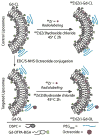
LP constructs and radiolabeling strategy: OCT was conjugated to preformed Gd-Control LPs (CL) resulting in targeted OCT-LP (OL). Both paramagnetic formulations were radiolabeled using 89Zr with a chelator-free approach.

Similar articles

Cited by

References

    1. Cherry SR. Multimodality imaging: beyond PET/CT and SPECT/CT. Semin Nucl Med. 2009;39(5):348–53. Epub 2009/08/04. - PMC - PubMed
    1. Catana C, Procissi D, Wu Y, Judenhofer MS, Qi J, Pichler BJ, et al. Simultaneous in vivo positron emission tomography and magnetic resonance imaging. Proc Natl Acad Sci U S A. 2008;105(10):3705–10. Epub 2008/03/06. - PMC - PubMed
    1. Bellin MF. MR contrast agents, the old and the new. Eur J Radiol. 2006;60(3):314–23. Epub 2006/09/29. - PubMed
    1. Shellock FG. MR imaging in patients with intraspinal bullets. J Magn Reson Imaging. 1999;10(1):107. Epub 1999/07/10. - PubMed
    1. Terreno E, Delli Castelli D, Cabella C, Dastru W, Sanino A, Stancanello J, et al. Paramagnetic liposomes as innovative contrast agents for magnetic resonance (MR) molecular imaging applications. Chem Biodivers. 2008;5(10):1901–12. Epub 2008/10/31. - PubMed

Publication types

LinkOut - more resources