

Primary hyperparathyroidism is associated with abnormal cortical and trabecular microstructure and reduced bone stiffness in postmenopausal women
- PMID: 23225022
- PMCID: PMC3631282
- DOI: 10.1002/jbmr.1841
Primary hyperparathyroidism is associated with abnormal cortical and trabecular microstructure and reduced bone stiffness in postmenopausal women
Abstract
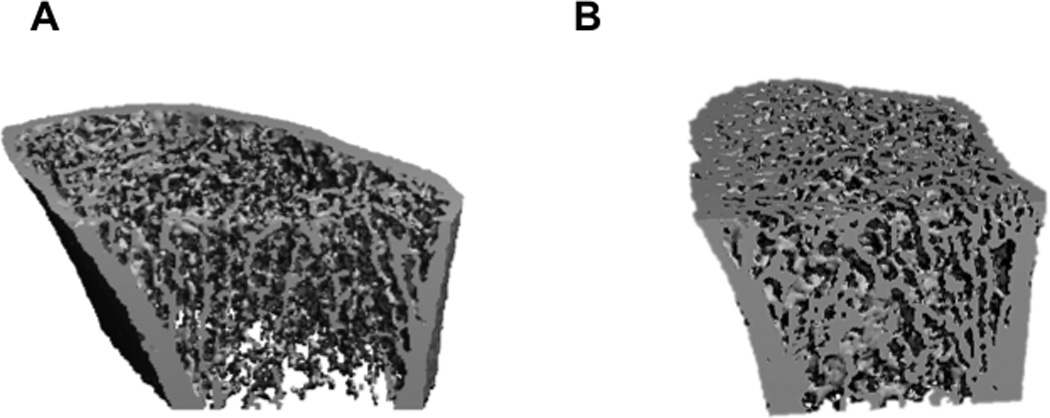
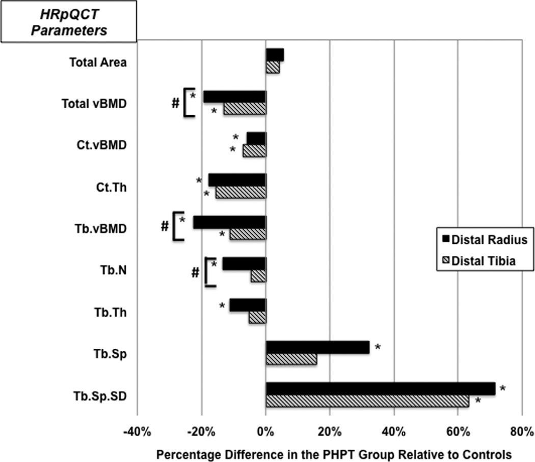
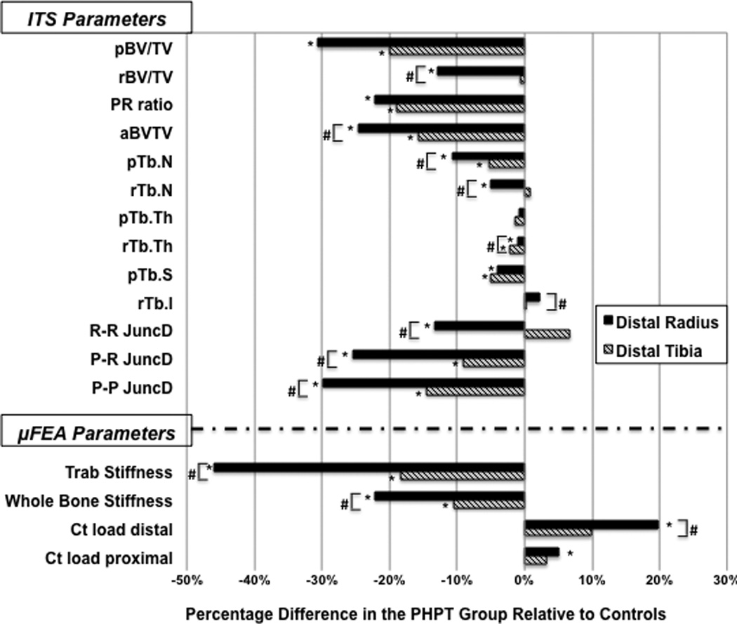
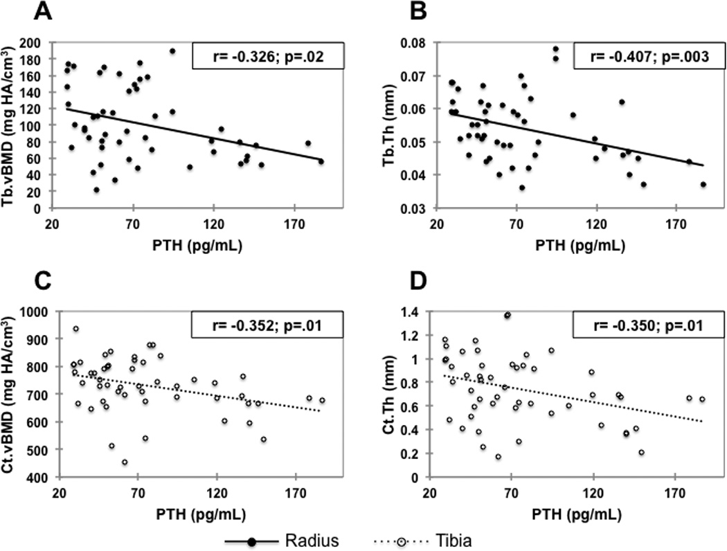
Typically, in the milder form of primary hyperparathyroidism (PHPT), now seen in most countries, bone density by dual-energy X-ray absorptiometry (DXA) and detailed analyses of iliac crest bone biopsies by histomorphometry and micro-computed tomography (µCT) show detrimental effects in cortical bone, whereas the trabecular site (lumbar spine by DXA) and the trabecular compartment (by bone biopsy) appear to be relatively well preserved. Despite these findings, fracture risk at both vertebral and nonvertebral sites is increased in PHPT. Emerging technologies, such as high-resolution peripheral quantitative computed tomography (HRpQCT), may provide additional insight into microstructural features at sites such as the forearm and tibia that have heretofore not been easily accessible. Using HRpQCT, we determined cortical and trabecular microstructure at the radius and tibia in 51 postmenopausal women with PHPT and 120 controls. Individual trabecula segmentation (ITS) and micro-finite element (µFE) analyses of the HRpQCT images were also performed to further understand how the abnormalities seen by HRpQCT might translate into effects on bone strength. Women with PHPT showed, at both sites, decreased volumetric densities at trabecular and cortical compartments, thinner cortices, and more widely spaced and heterogeneously distributed trabeculae. At the radius, trabeculae were thinner and fewer in PHPT. The radius was affected to a greater extent in the trabecular compartment than the tibia. ITS analyses revealed, at both sites, that plate-like trabeculae were depleted, with a resultant reduction in the plate/rod ratio. Microarchitectural abnormalities were evident by decreased plate-rod and plate-plate junctions at the radius and tibia, and rod-rod junctions at the radius. These trabecular and cortical abnormalities resulted in decreased whole-bone stiffness and trabecular stiffness. These results provide evidence that in PHPT, microstructural abnormalities are pervasive and not limited to the cortical compartment, which may help to account for increased global fracture risk in PHPT.
Copyright © 2013 American Society for Bone and Mineral Research.
Conflict of interest statement
Disclosures:
The authors state that they have no conflicts of interest
Figures
References
-
- Cope O. The study of hyperparathyroidism at the Massachusetts General Hospital. N Engl J Med. 1966;274(21):1174–1182. Epub 1966/05/26. - PubMed
-
- Silverberg SJ, Shane E, Jacobs TP, Siris E, Bilezikian JP. A 10-year prospective study of primary hyperparathyroidism with or without parathyroid surgery. N Engl J Med. 1999;341(17):1249–1255. Epub 1999/10/21. - PubMed
-
- Bilezikian JP, Brandi ML, Rubin M, Silverberg SJ. Primary hyperparathyroidism: new concepts in clinical, densitometric and biochemical features. J Intern Med. 2005;257(1):6–17. Epub 2004/12/21. [pii] 10.1111/j.1365-2796.2004.01422.x. - PubMed
-
- Silverberg SJ, Shane E, de la Cruz L, Dempster DW, Feldman F, Seldin D, et al. Skeletal disease in primary hyperparathyroidism. Journal of bone and mineral research : the official journal of the American Society for Bone and Mineral Research. 1989;4(3):283–291. Epub 1989/06/01. - PubMed
Publication types
MeSH terms
Grants and funding
- R01 AR051376/AR/NIAMS NIH HHS/United States
- DK32333/DK/NIDDK NIH HHS/United States
- K23 DK084337/DK/NIDDK NIH HHS/United States
- K24 AR052665/AR/NIAMS NIH HHS/United States
- UL1 RR024156/RR/NCRR NIH HHS/United States
- U01 AR055068/AR/NIAMS NIH HHS/United States
- K23 DK 084337/DK/NIDDK NIH HHS/United States
- R01 AR058004/AR/NIAMS NIH HHS/United States
- AR051376/AR/NIAMS NIH HHS/United States
- K23 DK095944/DK/NIDDK NIH HHS/United States
- K23 DK 095944/DK/NIDDK NIH HHS/United States
- R01 DK032333/DK/NIDDK NIH HHS/United States
- AR058004/AR/NIAMS NIH HHS/United States
- AR 055068/AR/NIAMS NIH HHS/United States
LinkOut - more resources
Full Text Sources
Other Literature Sources
