. 2013 Feb;73(2):309-15.
doi: 10.1002/ana.23793. Epub 2012 Dec 7.

Dopamine deficiency underlies learning deficits in neurofibromatosis-1 mice

Affiliations

Dopamine deficiency underlies learning deficits in neurofibromatosis-1 mice

Kelly A Diggs-Andrews et al. Ann Neurol. 2013 Feb.

Abstract

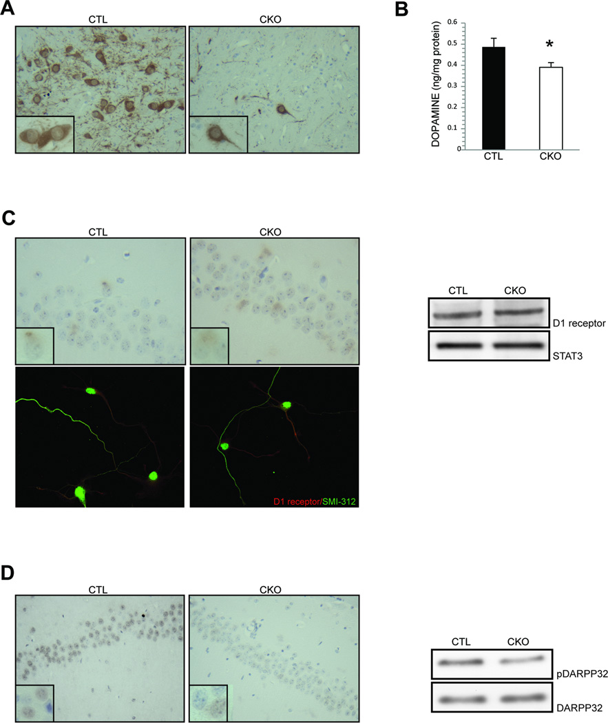
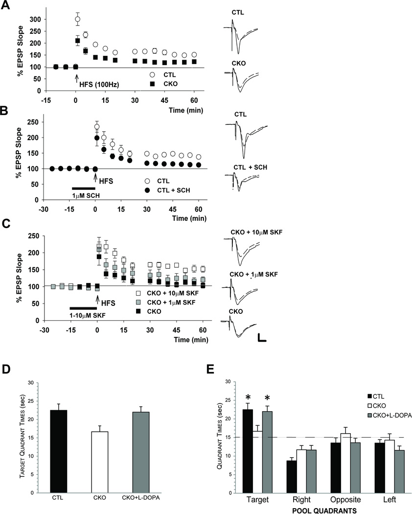
Children with neurofibromatosis type 1 (NF1) are prone to learning and behavioral abnormalities, including problems with spatial learning and attention. The molecular etiology for these deficits is unclear, as previous studies have implicated defective dopamine, cyclic adenosine monophosphate (cAMP), and Ras homeostasis. Using behavioral, electrophysiological, and primary culture, we now demonstrate that reduced dopamine signaling is responsible for cAMP-dependent defects in neuron function and learning. Collectively, these results establish defective dopaminergic function as a contributing factor underlying impaired spatial learning and memory in children and adults with NF1, and support the use of treatments that restore normal dopamine homeostasis for select individuals.


Figures

Figure 1
Figure 1. Reduced hippocampal dopamine signaling is observed in Nf1 mutant mice
(A) Reduced numbers of tyrosine hydroxylase (TH)-expressing neurons are detected in the ventral tegmental area (VTA) of CKO mice compared to controls. (B) Dopamine levels in the hippocampus of CKO mice are 20% lower than littermate controls (CTL). (C) Hippocampal D1 receptor expression is similar in CKO and control (CTL) mice in vivo and in vitro (1.0 v. 1.1 relative fold change). (D) Hippocampal DARPP32 phosphorylation is reduced by 3-fold in the CA1 region of CKO mice relative to littermate controls (CTL). Insets are representative 600x high-power field magnification images of individual cells. Asterisk denotes p<0.05.
Figure 2
Figure 2. Dopamine treatment rescues LTP and spatial learning deficits in Nf1 mutant mice
(A) In control (CTL) hippocampal slices, high frequency stimulation (HFS) induces long term potentiation (LTP). In contrast, reduced LTP is observed in CKO slices. (B) D1 receptor blockade with the antagonist SCH23390 (1 µM SCH) reduces LTP in control (CTL) slices. (C) D1 receptor activation by SKF38393 (SKF) in CKO slices induces a partial LTP rescue at 1 µM and a complete rescue at 10 µM. (D) During the second probe trial in the water maze, both control (CTL) and CKO mice treated with L-DOPA (50 mg/kg; CKO + L-DOPA) spent significantly more time in the target quadrant compared to the untreated CKO group (p=0.01, p=0.02, respectively). (E) Both control (CTL) and L-DOPA-treated (CKO + L-DOPA) mice spent more time in the target quadrant compared to each of the other quadrants (p<0.002 for target vs. other quadrant comparisons; dashed line represents chance) relative to untreated CKO mice (no spatial bias). Traces depict EPSPs before (dashed lines) and 60 min after HFS (solid lines). Scale = 1mV, 5 ms. Asterisks denote p<0.05.
Figure 3
Figure 3. Dopamine treatment restores normal hippocampal cAMP signaling in vivo and rescues Nf1+/− hippocampal neuron abnormalities in vitro
(A) L-DOPA treatment of CKO mice (CKO + L-DOPA) increases hippocampal cAMP levels to control (CTL) levels. (B) L-DOPA treatment of CKO mice (CKO + L-DOPA) increases hippocampal DARPP32 phosphorylation without affecting Ras activity or ERK phosphorylation. (C) Golgi staining of CKO hippocampal sections (CKO) reveals shorter axons and reduced neuronal branching (arborization) relative to littermate controls (CTL). (D) Primary cultured Nf1+/− hippocampal neurons have reduced axon lengths (SMI-312) and growth cone areas relative to wild-type (Nf1+/+) littermate controls. No differences in dendritic length were observed in Nf1+/− hippocampal neurons (MAP2). The neurite length and growth cone area deficits in Nf1+/− hippocampal neurons are rescued by dopamine (100 µM) treatment and D1 receptor activation (SKF38393, 1 µM). Scale bar = 50µm. Asterisks denote p<0.05.

References

    1. Mautner VF, Kluwe L, Thakker SD, Leark RA. Treatment of ADHD in neurofibromatosis type 1. Dev Med Child Neurol. 2002;44:164–170. - PubMed
    1. Hyman SL, Arthur Shores E, North KN. Learning disabilities in children with neurofibromatosis type 1: subtypes, cognitive profile, and attention-deficit-hyperactivity disorder. Dev Med Child Neurol. 2006;48:973–977. - PubMed
    1. Cui Y, Costa RM, Murphy GG, et al. Neurofibromin regulation of ERK signaling modulates GABA release and learning. Cell. 2008;135:549–560. - PMC - PubMed
    1. Li W, Cui Y, Kushner SA, et al. The HMG-CoA reductase inhibitor lovastatin reverses the learning and attention deficits in a mouse model of neurofibromatosis type 1. Curr Biol. 2005;15:1961–1967. - PubMed
    1. Brown JA, Emnett RJ, White CR, et al. Reduced striatal dopamine underlies the attention system dysfunction in neurofibromatosis-1 mutant mice. Hum Mol Genet. 2010;19:4515–4528. - PMC - PubMed

Publication types

MeSH terms