Comparative Study
. 2013 Jan;73(1):104-19.
doi: 10.1002/ana.23748. Epub 2012 Dec 7.

Amyloid-β oligomerization in Alzheimer dementia versus high-pathology controls

Affiliations
Comparative Study

Amyloid-β oligomerization in Alzheimer dementia versus high-pathology controls

Thomas J Esparza et al. Ann Neurol. 2013 Jan.

Abstract

Objective: Although amyloid-beta (Aβ) peptide deposition into insoluble plaques is a pathological hallmark of Alzheimer disease; soluble oligomeric Aβ has been hypothesized to more directly underlie impaired learning and memory in dementia of the Alzheimer type. However, the lack of a sensitive, specific, and quantitative assay for Aβ oligomers has hampered rigorous tests of this hypothesis.

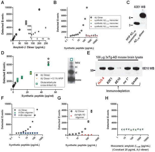
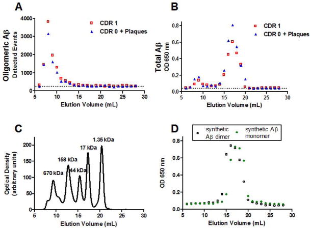
Methods: We developed a plate-based single molecule counting fluorescence immunoassay for oligomeric Aβ sensitive to low pg/ml concentrations of synthetic Aβ dimers using the same Aβ-specific monoclonal antibody to both capture and detect Aβ. The Aβ oligomer assay does not recognize monomeric Aβ, amyloid precursor protein, or other non-Aβ peptide oligomers.

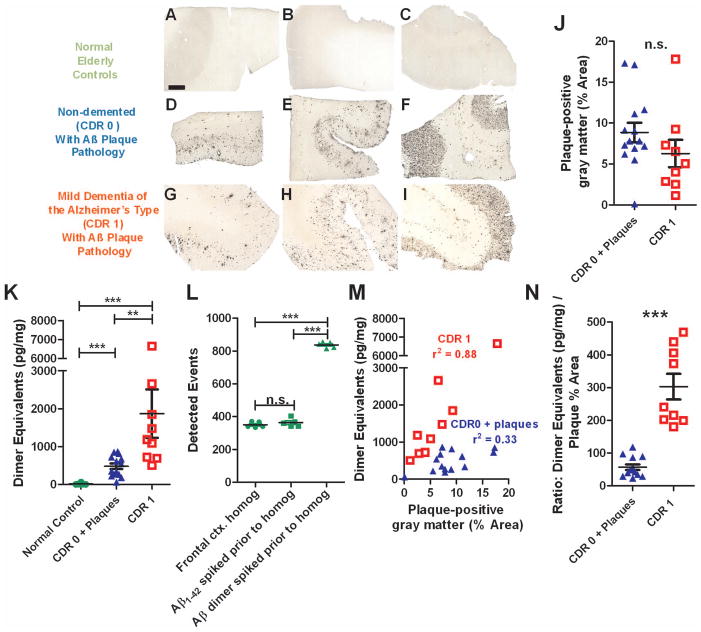
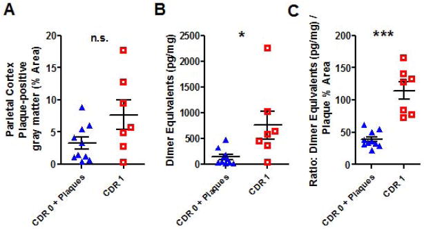
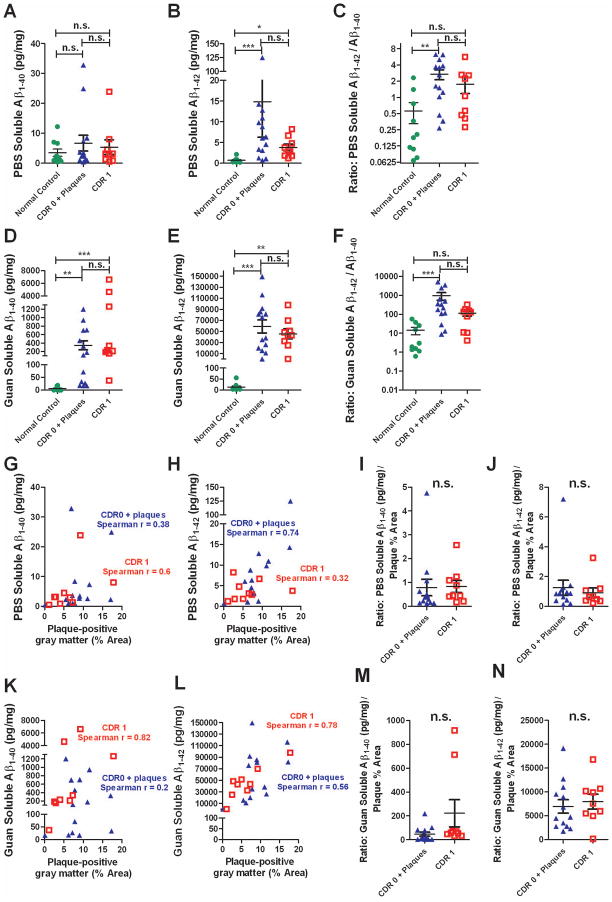
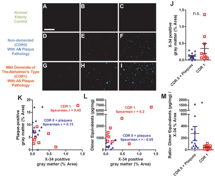
Results: Aβ oligomers were detected in aqueous cortical lysates from patients with dementia of the Alzheimer type and nondemented patients with Aβ plaque pathology. However, Aβ oligomer concentrations in demented patients' lysates were tightly correlated with Aβ plaque coverage (r = 0.88), but this relationship was weaker in those from nondemented patients (r = 0.30) despite equivalent Aβ plaque pathology. The ratio of Aβ oligomer levels to plaque density fully distinguished demented from nondemented patients, with no overlap between groups in this derived variable. Other Aβ and plaque measures did not distinguish demented from nondemented patients. Aβ oligomers were not detected in cerebrospinal fluid with this assay.

Interpretation: The results raise the intriguing hypothesis that the linkage between plaques and oligomers may be a key pathophysiological event underlying dementia of the Alzheimer type. This Aβ oligomer assay may be useful for many tests of the oligomer hypothesis.


Figures

Figure 1
Figure 1
Sensitivity and specificity characteristics of the Aβ oligomer assay. A. Standard curve demonstrating a monotonic relationship between detected events and the concentration of synthetic Aβ1-40Ser26Cys dimer. Inset: concentrations of Aβdimer as low as 3.125 pg/ml were detectible above background. B. Specificity for Aβ dimers over Aβmonomers. (Note the log scale of the x-axis) C. Western blot demonstrating that the Aβ monomer preparation did not contain dimer and the Aβ1-40Ser26Cys dimer preparation did not contain Aβ monomer. D. Sensitivity of the assay to another type of synthetic Aβ oligomers, prepared using aggregated wild-type sequence Aβ cross-linked with glutaradehyde. Right: Western blot demonstrating a mixture of monomers, dimers, trimers and tetramers in this preparation. E. Immunodepletion experiment demonstrating that HJ3.4, the antibody used in the Aβ oligomer assay, does not bind APP in solution. Controls: 82E1 is known to require a free amino terminus of Aβ to bind, and therefore does not recognize APP, whereas 6E10 is known to bind both APP and Aβ (Reproduced from Tran et al. 2011). F. Specificity for Aβ dimers over aggregated A-Dan and A-Bri, two non-Aβ peptides that readily oligomerize. (Note the log scale of the x-axis) G. Specificity for full length Aβ dimers over N-terminally truncated, pyroglutamate modified Aβ oligomers. H. Sensitivity to Aβ dimers is unchanged in the presence of high concentrations of Aβ monomers.
Figure 2
Figure 2
Oligomerization of Aβ is tightly linked to plaque density in dementia of the Alzheimer type but not in high pathology elderly controls. A–C. Aβ immunohistochemistry using a polyclonal rabbit anti-pan-Aβ on frontal cortex sections from normal elderly controls demonstrates the absence of plaque pathology. Scale bar: 1 mm applies to panels A–I. D–F. Aβ plaque pathology in frontal cortex sections from non-demented elderly subjects (CDR 0). G–I. Aβ plaque pathology in frontal cortex sections from elderly subjects with mild dementia of the Alzheimer’s type (CDR 1). J. Gray matter coverage by Aβ plaque pathology was not different in the non-demented elderly subjects with plaques (CDR 0 + plaques) vs. subjects with mild dementia of the Alzheimer’s type (CDR 1). K. Aβ oligomer levels in PBS-soluble frontal cortical homogenates. (** p=0.0023, *** p=0.0003, Mann Whitney U tests). Oligomer levels expressed as pg dimer equivalents per mg total protein in homogenates. L. Control for artifactual oligomerization of monomeric Aβ during homogenization and analysis. The addition of 1000 pg/ml of monomeric Aβ1-42 spiked into the homogenization buffer along with brain tissue from normal control subjects did not result in any detectible oligomer signal. The presence of brain homogenate did not obscure or block oligomer detection, as spiking in Aβdimer resulted in the expected elevation in signal (*** p=0.0001, 1-way ANOVA). M. Correlations between Aβ oligomer levels (y-axis) and gray matter Aβ plaque pathology coverage (x-axis). r2 values represent Pearson product moment correlations. N. Ratio of Aβ oligomer levels to Aβ plaque pathology coverage was higher in subjects with mild dementia of the Alzheimer’s type (CDR 1) vs non-demented elderly subjects with plaques (CDR 0 + plaques). There was no overlap between groups (***p=0.0001, Mann Whitney U test).
Figure 3
Figure 3
Similar relationship between Aβ oligomerization and plaque pathology in parietal cortex. A. Parietal gray matter coverage by Aβ plaque pathology was not different in the non-demented elderly subjects with plaques (CDR 0 + plaques) vs. subjects with mild dementia of the Alzheimer’s type (CDR 1). B. Aβ oligomer levels in PBS-soluble parietal cortical homogenates. (* p=0.014, Mann Whitney U test). C. Ratio of Aβ oligomer levels to Aβ plaque pathology coverage was higher in subjects with mild dementia of the Alzheimer’s type (CDR 1) vs non-demented elderly subjects with plaques (CDR 0 + plaques). As in frontal cortex, there was no overlap between groups (***p=0.0001, Mann Whitney U test).
Figure 4
Figure 4
Assessments based on overall Aβ levels did not distinguish tissue from patients with mild dementia of the Alzheimer type (CDR 1) vs. non-demented elderly patients with plaque pathology (CDR 0 + plaque). A. No difference between groups in overall PBS-soluble Aβ1-40 levels, as measured using a standard sandwich ELISA. Data expressed as pg of Aβ per mg total protein. B. Overall PBS-soluble Aβ1-42 levels were not different in the CDR 0 + plaque vs. CDR 1 group, though levels in both groups were higher than in the normal control group (*p < 0.05, *** p < 0.001, Kruskall Wallis ANOVA with Dunn’s post-hoc test). C. Ratio of overall PBS-soluble Aβ1-42 levels to overall PBS-soluble Aβ1-40 levels did not distinguish CDR 0 + plaque vs. CDR 1 groups. The CDR 0 + plaque group had higher ratios than the control group (**p < 0.01). D–E. Overall Guanidine-soluble Aβ1-40 levels and Aβ1-42 levels were not different in the CDR 0 + plaque vs. CDR 1 group. Levels in both groups were higher than in the normal control group for both measures. F. Ratio of overall Guanidine-soluble Aβ1-42 levels to overall PBS-soluble Aβ1-40 levels did not distinguish CDR 0 + plaque vs. CDR 1 groups. The CDR 0 + plaque group had higher ratios than the control group (*** p < 0.001). G–H. Correlations between overall PBS-soluble Aβ1-40 and Aβ1-42 levels vs. plaque-positive gray matter. The correlation was only significant (p = 0.0023) for PBS-soluble Aβ1-42 vs. plaque area in the CDR 0 + plaque group. I–J. Ratios of overall PBS-soluble Aβ1-40 and Aβ1-42 levels to plaque-positive gray matter area coverage did not distinguish CDR 0 + plaque vs. CDR 1 groups (n. s. not significant, Mann-Whitney U Tests). K–L. Significant correlations between overall Guan-soluble Aβ1-40 and Aβ1-42 levels vs. plaque-positive gray matter. (p = 0.01 for Guan-soluble Aβ1-40 vs. plaque area in the CDR 1 group, p = 0.02 for Guan-soluble Aβ1-42 vs. plaque area in the CDR 0 + plaque group, p = 0.04 for Guan-soluble Aβ1-42 vs. plaque area in the CDR 1 group). M–N. Ratios of overall Guan-soluble Aβ1-40 and Aβ1-42 levels to plaque-positive gray matter area coverage did not distinguish CDR 0 + plaque vs. CDR 1 groups (n. s. not significant, Mann-Whitney U Tests).
Figure 5
Figure 5
Assessments based on X-34 staining of fibrillar plaque pathology did not distinguish between tissue from patients with mild dementia of the Alzheimer type (CDR 1) vs. non-demented elderly patients with plaque pathology (CDR 0 + plaque). A–I: Exemplar images of X-34 staining in frontal cortex sections. Scale bar = 1 mm. J. Gray matter coverage by X-34 in CDR 0 + plaques group vs. CDR 1 group (n.s., Mann Whitney U test). K. Correlations between overall Aβ plaque coverage vs. X-34 positive fibrillar plaque coverage. (Spearman r = 0.65, p = 0.06 for the CDR 1 group). L. Correlations between Aβ oligomer levels (dimer equivalents) and gray matter X-34 positive fibrillar plaque pathology coverage. M. Ratio of Aβ oligomer levels (dimer equivalents) to X-34 positive fibrillar plaque pathology coverage was higher in the CDR 0 + plaque group (p = 0.02, Mann Whitney U test). However, there was substantial overlap between groups.
Figure 6
Figure 6
Size exclusion chromatography indicated that Aβ oligomers were primarily high molecular weight both in non-demented patients with Aβ plaque pathology (CDR 0 + Plaques) and patients with mild dementia of the Alzheimer type (CDR 1). A. Plate-based fluorescence immunoassay for Aβ oligomers, reported as detected events, for serial fractions eluted with PBS off of a Superdex 200 10/300 GL column: larger molecules elute in earlier fractions, and smaller molecules elute in later fractions. Aβ oligomers eluted in fractions 7–10 for both samples. Dashed line indicates background levels of detected events. B. Total Aβ assessed by indirect ELISA in the same fractions. Total Aβ eluted in 2 peaks, a high molecular weight peak in fractions 7–10 corresponding to oligomeric Aβ shown in panels A, and a lower molecular weight peak in fractions 15–19 likely corresponding to monomeric Aβ. C. Globular protein size standards, run on the same column under the same conditions. D. Synthetic Aβ dimers run on the same column under the same conditions eluted in fractions 14–19, and synthetic Aβ monomers eluted in fractions 15–19, as assessed by total Aβ indirect ELISA.

References

    1. Price JL, Morris JC. Tangles and plaques in nondemented aging and “preclinical” Alzheimer’s disease. Annals of neurology. 1999 Mar;45(3):358–68. - PubMed
    1. Aizenstein HJ, Nebes RD, Saxton JA, et al. Frequent amyloid deposition without significant cognitive impairment among the elderly. Archives of neurology. 2008 Nov;65(11):1509–17. - PMC - PubMed
    1. Hulette CM, Welsh-Bohmer KA, Murray MG, Saunders AM, Mash DC, McIntyre LM. Neuropathological and neuropsychological changes in “normal” aging: evidence for preclinical Alzheimer disease in cognitively normal individuals. Journal of neuropathology and experimental neurology. 1998 Dec;57(12):1168–74. - PubMed
    1. Katzman R, Terry R, DeTeresa R, et al. Clinical, pathological, and neurochemical changes in dementia: a subgroup with preserved mental status and numerous neocortical plaques. Annals of neurology. 1988 Feb;23(2):138–44. - PubMed
    1. Selkoe DJ. Soluble oligomers of the amyloid beta-protein impair synaptic plasticity and behavior. Behav Brain Res. 2008 Sep 1;192(1):106–13. - PMC - PubMed

Publication types

Substances