Case Reports
. 2013 Jan;73(1):120-8.
doi: 10.1002/ana.23756. Epub 2012 Dec 7.

Encephalitis and antibodies to dipeptidyl-peptidase-like protein-6, a subunit of Kv4.2 potassium channels

Affiliations
Case Reports

Encephalitis and antibodies to dipeptidyl-peptidase-like protein-6, a subunit of Kv4.2 potassium channels

Anna Boronat et al. Ann Neurol. 2013 Jan.

Abstract

Objective: To report a novel cell surface autoantigen of encephalitis that is a critical regulatory subunit of the Kv4.2 potassium channels.

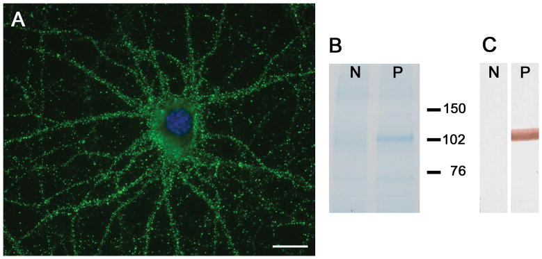
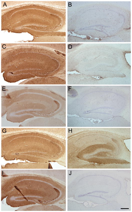
Methods: Four patients with encephalitis of unclear etiology and antibodies with a similar pattern of neuropil brain immunostaining were selected for autoantigen characterization. Techniques included immunoprecipitation, mass spectrometry, cell-base experiments with Kv4.2 and several dipeptidyl-peptidase-like protein-6 (DPPX) plasmid constructs, and comparative brain immunostaining of wild-type and DPPX-null mice.

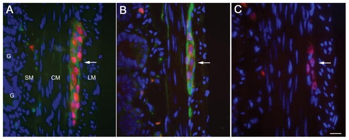
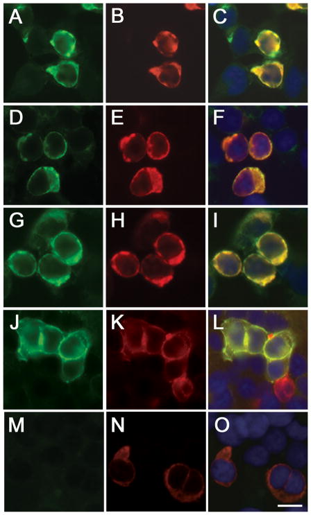
Results: Immunoprecipitation studies identified DPPX as the target autoantigen. A cell-based assay confirmed that all 4 patients, but not 210 controls, had DPPX antibodies. Symptoms included agitation, confusion, myoclonus, tremor, and seizures (1 case with prominent startle response). All patients had pleocytosis, and 3 had severe prodromal diarrhea of unknown etiology. Given that DPPX tunes up the Kv4.2 potassium channels (involved in somatodendritic signal integration and attenuation of dendritic back-propagation of action potentials), we determined the epitope distribution in DPPX, DPP10 (a protein homologous to DPPX), and Kv4.2. Patients' antibodies were found to be specific for DPPX, without reacting with DPP10 or Kv4.2. The unexplained diarrhea led to a demonstration of a robust expression of DPPX in the myenteric plexus, which strongly reacted with patients' antibodies. The course of neuropsychiatric symptoms was prolonged and often associated with relapses during decreasing immunotherapy. Long-term follow-up showed substantial improvement in 3 patients (1 was lost to follow-up).

Interpretation: Antibodies to DPPX are associated with a protracted encephalitis characterized by central nervous system hyperexcitability (agitation, myoclonus, tremor, seizures), pleocytosis, and frequent diarrhea at symptom onset. The disorder is potentially treatable with immunotherapy.


Figures

Figure 1
Figure 1. Immunoprecipitation of DPPX
In cultures of dissociated rat hippocampal neurons, patients’ antibodies showed intense reactivity with the neuronal cell surface (A), bar = 10 μm. Immunoprecipitation of the antigen with serum of the index case is shown in B, where the precipitated proteins were run in a gel and subsequently stained with EZblue. Note that patient’s antibodies precipitated a protein (band close to 102 kDa in lane P), which was excised from the gel and analyzed by mass spectrometry, demonstrating sequences of DPPX. Lane N is the precipitate obtained from control serum. Immunoblot of these proteins with a rabbit polyclonal antibody against DPPX (1:1000, developed by BR) confirmed that the band corresponded to DPPX (C).
Figure 2
Figure 2. Expression of DPPX in myenteric plexus
Transverse section of small bowel of rat showing the longitudinal muscular layer (LM), circular muscular layer (CM), submucosal layer (SM), and glans (G). The myenteric plexus (Plex) is revealed as clusters of large neurons between the two muscular layers. In the 3 panels (A–C) the nuclei of the neurons (red) was labeled with anti-Hu (a highly specific neuronal marker). Panel A, shows in green the DPPX immunostaining using a rabbit polyclonal antibody (1:1000, developed by BD); panel B shows the DPPX reactivity of serum from one of the patients with encephalitis, and panel C shows the lack of reactivity of serum from a healthy subject. Note that DPPX is predominantly expressed in the cytoplasm-membrane of the large clustered neurons of the myenteric plexus, and is also detected in a fine longitudinal pattern in CM and SM where the submucosal plexus is located. Bar = 20μm.
Figure 3
Figure 3. Analysis of DPPX antibodies using a cell-based assay
HEK 293 cells expressing DPPX-L immunostained with patients’ serum (A, D, G, J) and a mouse monoclonal antibody against DPPX (B, E, H, K). The merged reactivities are shown in the corresponding panels (C, F, I, L). Similar studies comparing the serum of a healthy individual and the DPPX monoclonal antibody are shown in M and N, and the merged reactivities in O. Note that patient’s antibodies immunoreact with cells expressing DPPX. Bar = 10 μm.
Figure 4
Figure 4. Comparison of patients’ serum reactivity using brain from DPPX-null mutants and wild type mice
The reactivity of patients’ serum with the hippocampus of wild type mice is shown in A, C, E and G. The reactivity of a rabbit polyclonal DPPX antibody with the hippocampus of wild-type mice is shown in I. Panels on the right side show the results of a similar experiment but using the hippocampus of DPPX-null mice. Note that the reactivities of the sera of the first three patients (cases 1, 2, and 3 of Table 1) and the rabbit polyclonal DPPX antibody are abrogated in the hippocampus of DPPX-null mice (panels B, D, F, J). Patient 4, not included in the table (panels G and H) showed remaining reactivity with the hippocampus of DPPX-null mice indicating that this patient had two antibodies, one against DPPX and the other against an unknown antigen. Bar = 200 μm.

References

    1. Lancaster E, Martinez-Hernandez E, Dalmau J. Encephalitis and antibodies to synaptic and neuronal cell surface proteins. Neurology. 2011;77:179–189. - PMC - PubMed
    1. Frechette ES, Zhou L, Galetta SL, Chen L, Dalmau J. Prolonged follow-up and CSF antibody titers in a patient with anti-NMDA receptor encephalitis. Neurology. 2011;76:S64–S66. - PubMed
    1. Gable MS, Sheriff H, Dalmau J, Tilley DH, Glaser CA. The Frequency of Autoimmune N-Methyl-D-Aspartate Receptor Encephalitis Surpasses That of Individual Viral Etiologies in Young Individuals Enrolled in the California Encephalitis Project. Clin Infect Dis. 2012 - PMC - PubMed
    1. Gable MS, Gavali S, Radner A, Tilley DH, Lee B, Dyner L, et al. Anti-NMDA receptor encephalitis: report of ten cases and comparison with viral encephalitis. Eur J Clin Microbiol Infect Dis. 2009;28:1421–1429. - PMC - PubMed
    1. Ances BM, Vitaliani R, Taylor RA, Liebeskind DS, Voloschin A, Houghton DJ, et al. Treatment-responsive limbic encephalitis identified by neuropil antibodies: MRI and PET correlates. Brain. 2005;128:1764–1777. - PMC - PubMed

Publication types

MeSH terms