. 2014 Mar;35(3):819-30.
doi: 10.1002/hbm.22216. Epub 2012 Dec 8.

The burden of microstructural damage modulates cortical activation in elderly subjects with MCI and leuko-araiosis. A DTI and fMRI study

Collaborators, Affiliations

The burden of microstructural damage modulates cortical activation in elderly subjects with MCI and leuko-araiosis. A DTI and fMRI study

Mario Mascalchi et al. Hum Brain Mapp. 2014 Mar.

Abstract

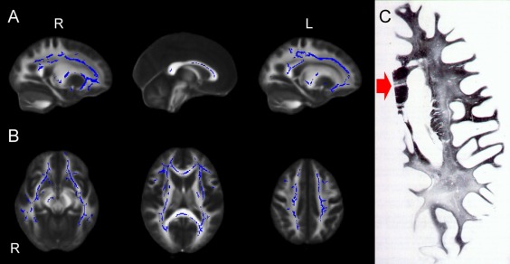
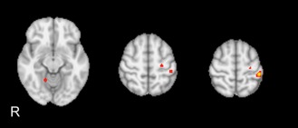
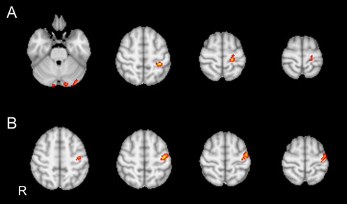
The term leuko-araiosis (LA) describes a common chronic affection of the cerebral white matter (WM) in the elderly due to small vessel disease with variable clinical correlates. To explore whether severity of LA entails some adaptive reorganization in the cerebral cortex we evaluated with functional MRI (fMRI) the cortical activation pattern during a simple motor task in 60 subjects with mild cognitive impairment and moderate or severe (moderate-to-severe LA group, n = 46) and mild (mild LA group, n = 14) LA extension on visual rating. The microstructural damage associated with LA was measured on diffusion tensor data by computation of the mean diffusivity (MD) of the cerebral WM and by applying tract based spatial statistics (TBSS). Subjects were examined with fMRI during continuous tapping of the right dominant hand with task performance measurement. Moderate-to-severe LA group showed hyperactivation of left primary sensorimotor cortex (SM1) and right cerebellum. Regression analyses using the individual median of WM MD as explanatory variable revealed a posterior shift of activation within the left SM1 and hyperactivation of the left SMA and paracentral lobule and of the bilateral cerebellar crus. These data indicate that brain activation is modulated by increasing severity of LA with a local remapping within the SM1 and increased activity in ipsilateral nonprimary sensorimotor cortex and bilateral cerebellum. These potentially adaptive changes as well lack of contralateral cerebral hemisphere hyperactivation are in line with sparing of the U fibers and brainstem and cerebellar WM tracts and the emerging microstructual damage of the corpus callosum revealed by TBSS with increasing severity of LA.

Keywords: diffusion tensor; functional MRI; leuko-araiosis; motor function.


Figures

Figure 1
Figure 1
(A–C) Sagittal (A) and axial (B) view of the results of voxel‐based analysis of MD maps by using TBSS. Blue shows regions where MD correlates significantly with LA visual score. All results are adjusted for age and gender. Permutation‐based nonparametric inference (10,000 permutations) including full correction for multiple comparison over space was performed using threshold‐free cluster enhancement (TFCE). An increased MD of the long cerebral WM tracts and of the transcallosal fibers with relative sparing of the U fibers is observed with increasing LA visual score. The WM tracts in the brainstem and cerebellum are essentially preserved except for a small cluster of increased MD in the right cerebral peduncle. (C) Woelke stain for myelin in a case of Binswanger's disease shows diffuse discoloration of the left cerebral white matter with relative sparing of the U fibers. Note the linear discoloration in the genu of the corpus callosum (red arrow) consistent with selective trancallosal tract degeneration (reprinted with permission from Mascalchi et al. Can J Neurol Sci 1989;16:214–218).
Figure 2
Figure 2
Between‐group statistical activation map. The moderate‐to‐severe LA group shows significant (Z > 10, P = 0.001 corrected) hyperactivation of left precentral and postcentral gyrus and right cerebellar lobule 5 as compared to mild LA group. No area of hypoactivation was observed in the moderate‐to‐severe LA group.
Figure 3
Figure 3
(A, B) Regression analyses in 60 subjects obtained by introducing median of the WM MD as a further explanatory variable in the GLM design matrix. (A) Activation significantly (Z > 8.5, P = 0.001 corrected) increases with increasing MD in the left pre‐ and postcentral gyrus extending to the left SMA and left paracentral lobule, respectively, and in the bilateral cerebellar crus. (B) Activation increases with decreasing MD in a single circumscribed area in left precentral gyrus.

Similar articles

Cited by

References

    1. Arsigny V, Fillard P, Pennec X, Ayache N (2006): Log‐euclidean metrics for fast and simple calculus on diffusion tensors. Magn Reson Med 56:411–421. - PubMed
    1. Avants BB, Yushkevich P, Pluta J, Minkoff D, Korczykowski M, Detre J, Gee JC (2010): The optimal template effect in hippocampus studies of diseased populations. NeuroImage 49:2457–2466. - PMC - PubMed
    1. Avants BB, Tustison NJ, Song G, Cook PA, Klein A, Gee JC (2011): A reproducible evaluation of ANTs similarity metric performance in brain image registration. NeuroImage 54:2033–2044. - PMC - PubMed
    1. Beckmann C, Jenkinson M, Smith SM (2003): General multi‐level linear modelling for group analysis in FMRI. NeuroImage 20:1052–1063. - PubMed
    1. Brickman AM, Siedlecki KL, Muraskin J, Manly JJ, Luchsinger JA, Yeung LK, Brown TR, De Carli C, Stern Y (2011): White matter hyperintensities and cognition: Testing the reserve hypothesis. Neurobiol Aging 32:1588–1598. - PMC - PubMed

Publication types

MeSH terms