. 2012 Nov;14(11):1087-96.
doi: 10.1593/neo.121342.

Long-range transcriptome sequencing reveals cancer cell growth regulatory chimeric mRNA

Affiliations

Long-range transcriptome sequencing reveals cancer cell growth regulatory chimeric mRNA

Roberto Plebani et al. Neoplasia. 2012 Nov.

Abstract

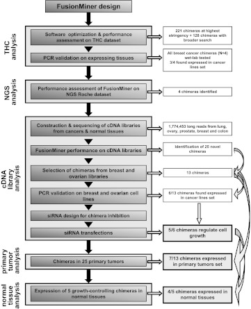
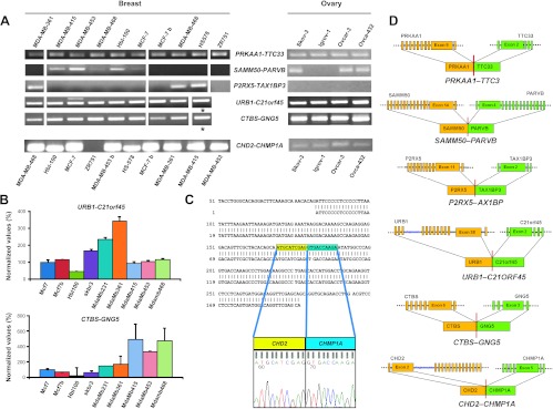
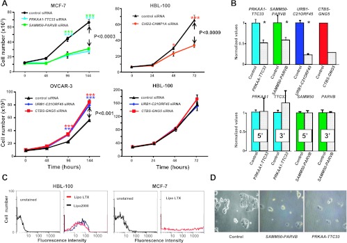
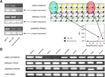
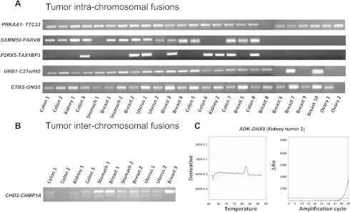
mRNA chimeras from chromosomal translocations often play a role as transforming oncogenes. However, cancer transcriptomes also contain mRNA chimeras that may play a role in tumor development, which arise as transcriptional or post-transcriptional events. To identify such chimeras, we developed a deterministic screening strategy for long-range sequence analysis. High-throughput, long-read sequencing was then performed on cDNA libraries from major tumor histotypes and corresponding normal tissues. These analyses led to the identification of 378 chimeras, with an unexpectedly high frequency of expression (≈2 x 10(-5) of all mRNA). Functional assays in breast and ovarian cancer cell lines showed that a large fraction of mRNA chimeras regulates cell replication. Strikingly, chimeras were shown to include both positive and negative regulators of cell growth, which functioned as such in a cell-type-specific manner. Replication-controlling chimeras were found to be expressed by most cancers from breast, ovary, colon, uterus, kidney, lung, and stomach, suggesting a widespread role in tumor development.


Figures

Figure 1
Figure 1
Flow diagram of chimera identification and validation steps.
Figure 2
Figure 2
Chimeric mRNA expression in cancer cell lines. (A) Expression of chimeras discovered from tumor and normal tissue library sequencing; agarose electrophoresis of nested or real-time PCR products. Breast and ovarian cancer cell lines are indicated; *SK-BR-3. (B) URB1-C21ORF45 (top) and CTBS-GNG5 (bottom) expression in breast cancer cell lines by quantitative RT-PCR; results are expressed as percent values (MCF-7 = 100); three replica samples were analyzed per data point. Bars, SD. (C) CHD2-CHMP1A. Sequence of the PCR amplicon versus that of the chimera isolated from the breast library. (D) Structure of validated chimeric mRNA; 5′ partners (orange) and 3′ partners (green) are shown; exon junctions are indicated.
Figure 3
Figure 3
Cell growth modulation by chimeras. (A) Control (Table S10) and chimera-targeting siRNA are color coded. (Top, left) MCF-7; PRKAA-TTC33 (cyan), SAMM50-PARVB (green), control (black). (Top, right) HBL-100; CHD2-CHMP1A (orange), control (black). (Bottom, left) OVCAR-3; URB1-C21ORF45 (blue), CTBS-GNG5 (red). (Bottom, right) HBL-100 treated with siRNA for chimeras from ovarian libraries; URB1-C21ORF45 (blue), CTBS-GNG5 (red). Bars, SD. Brackets, P value of two-way analysis of variance; Bonferroni t test significance: *P ≤ .05; ***P ≤ .001. (B) Real-time PCR of siRNA-transfected cells. (Top) Chimeric RNA (left to right: PRKAA-TTC33 and SAMM50-PARVB in MCF-7; URB1-C21ORF45 and CTBS-GNG5 in OVCAR-3). (Bottom) Single-partner RNA expression after the indicated siRNA treatment (left to right: PRKAA-TTC33 and SAMM50-PARVB in MCF-7). (C) Flow cytometry analysis of transfected HBL-100 and MCF7 cells; YFP was used as a transfection efficiency benchmark. (Left) HBL-100; LTX (red) or Lipo-2000 (blue). (Right) MCF-7; LTX transfection. (D) MCF-7 cell growth blockade after PRKAA-TTC33-targeted or SAMM50-PARVB-targeted siRNA treatment (day 6 after transfection).
Figure 4
Figure 4
Expression of mRNA chimeras in normal tissues. (A) (Left) Agarose electrophoresis analysis of real-time PCR products (Sybr Green real-time PCR). (Right) Real-time PCR analysis of the CHD2-CHMP1A chimeric mRNA in normal tissues (PrimeTime real-time PCR). HBL-100 cell cDNA was used as a positive control. (Bottom) Amplification plots of CHD2-CHMP1A mRNA in the HBL-100 breast cancer cell line (blue) and normal breast tissue (red). CHD2-CHMP1A (top lanes) and GAPDH (bottom lanes) were amplified in duplicate. Green triangles, successful amplification; red triangles, no amplification. (B) Agarose electrophoresis of nested PCR products of PRKAA1-TTC33, SAMM50-PARVB, CTBS-GNG5, and URB1-C21orf45 in normal tissues.
Figure 5
Figure 5
Expression of mRNA chimeras in primary tumors. (A, B) Agarose electrophoresis analysis of amplification products. Tumor origin and sample numbers are indicated. (A) Intrachromosomal chimeras as analyzed by real-time PCR. PRKAA1-TTC33 and CTBS-GNG5 were diagnosed in all 25 tumors; SAMM50-PARVB chimeric was found in 15 tumors, P2RX5-TAX1BP3 in 8 tumors, and URB1-C21orf45 in 21 tumors. (B) PCR amplicons of the interchromosomal CHD2-CHMP1A chimera. (C) Interchromosomal ADK-DHX8 chimeric. Melting temperature and real-time amplification curves.

References

    1. Mitelman F, Johansson B, Mertens F. The impact of translocations and gene fusions on cancer causation. Nat Rev Cancer. 2007;7:233–245. - PubMed
    1. Mercer TR, Dinger ME, Mattick JS. Long non-coding RNAs: insights into functions. Nat Rev Genet. 2009;10:155–159. - PubMed
    1. Tomlins SA, Rhodes DR, Perner S, Dhanasekaran SM, Mehra R, Sun XW, Varambally S, Cao X, Tchinda J, Kuefer R, et al. Recurrent fusion of TMPRSS2 and ETS transcription factor genes in prostate cancer. Science. 2005;310:644–648. - PubMed
    1. Rickman DS, Pflueger D, Moss B, VanDoren VE, Chen CX, de la Taille A, Kuefer R, Tewari AK, Setlur SR, Demichelis F, et al. SLC45A3ELK4 is a novel and frequent erythroblast transformation-specific fusion transcript in prostate cancer. Cancer Res. 2009;69:2734–2738. - PMC - PubMed
    1. Soda M, Choi YL, Enomoto M, Takada S, Yamashita Y, Ishikawa S, Fujiwara S, Watanabe H, Kurashina K, Hatanaka H, et al. Identification of the transforming EML4-ALK fusion gene in non-small-cell lung cancer. Nature. 2007;448:561–566. - PubMed

Supplemental References

    1. Wilming LG, Gilbert JG, Howe K, Trevanion S, Hubbard T, Harrow JL. The vertebrate genome annotation (Vega) database. Nucleic Acids Res. 2008;36:D753–D760. - PMC - PubMed
    1. Pruitt KD, Tatusova T, Klimke W, Maglott DR. NCBI Reference Sequences: current status, policy and new initiatives. Nucleic Acids Res. 2009;37:D32–D36. - PMC - PubMed
    1. Karolchik D, Kuhn RM, Baertsch R, Barber GP, Clawson H, Diekhans M, Giardine B, Harte RA, Hinrichs AS, Hsu F, et al. The UCSC Genome Browser Database: 2008 update. Nucleic Acids Res. 2008;36:D773–D779. - PMC - PubMed
    1. Romani A, Guerra M, Trerotola M, Alberti S. Detection and analysis of spliced chimeric mRNAs in sequence databanks. Nucleic Acids Res. 2003;31:1–8. - PMC - PubMed
    1. Calin GA, Sevignani C, Dumitru CD, Hyslop T, Noch E, Yendamuri S, Shimizu M, Rattan S, Bullrich F, Negrini M, et al. Human microRNA genes are frequently located at fragile sites and genomic regions involved in cancers. Proc Natl Acad Sci U S A. 2004;101:2999–3004. - PMC - PubMed

Publication types

LinkOut - more resources