. 2012;7(12):e49974.
doi: 10.1371/journal.pone.0049974. Epub 2012 Dec 3.

E4 antibodies facilitate detection and type-assignment of active HPV infection in cervical disease

Affiliations

E4 antibodies facilitate detection and type-assignment of active HPV infection in cervical disease

Heather Griffin et al. PLoS One. 2012.

Abstract

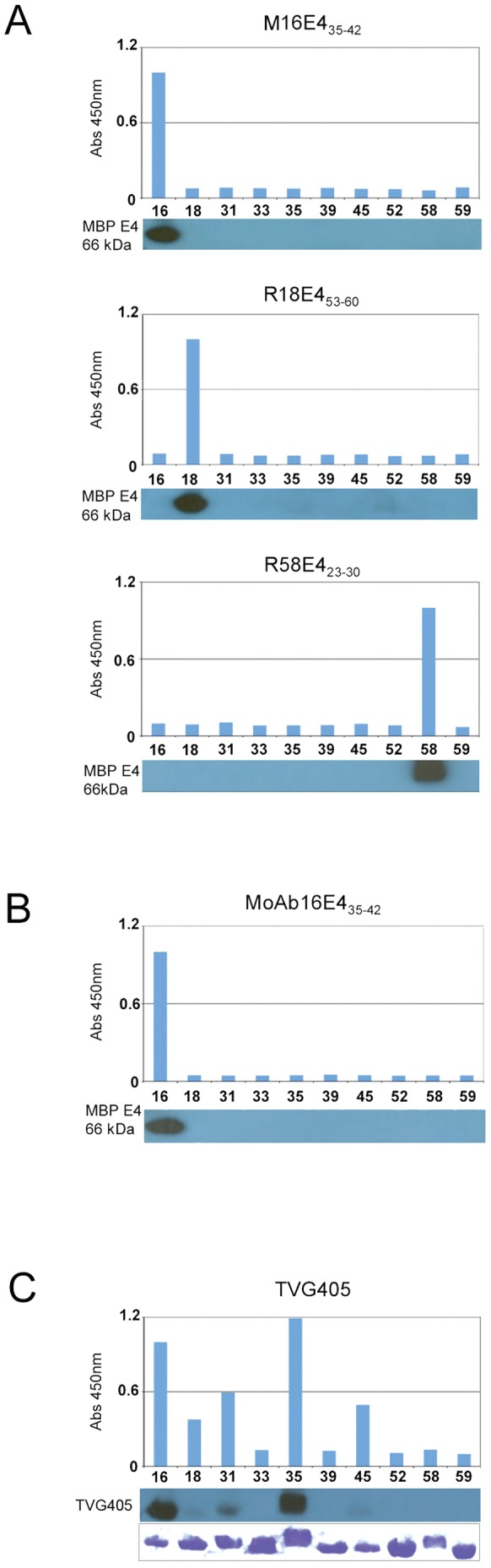
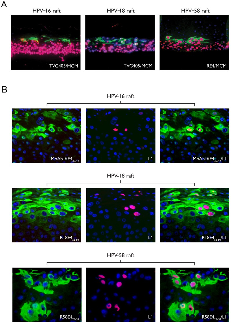
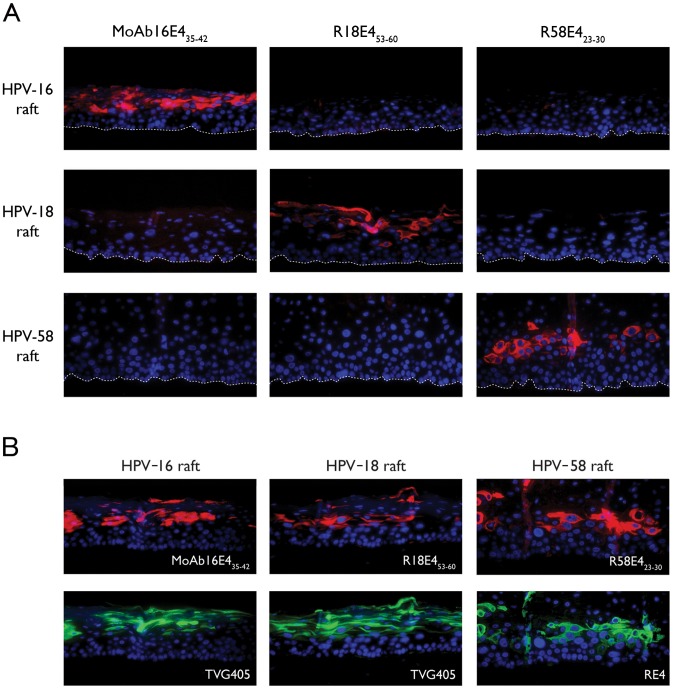
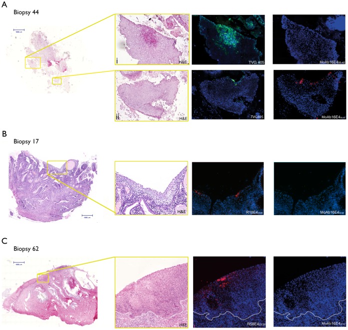
High-risk human papillomavirus (HPV) infections are the cause of nearly all cases of cervical cancer. Although the detection of HPV DNA has proved useful in cervical diagnosis, it does not necessarily predict disease presence or severity, and cannot conclusively identify the causative type when multiple HPVs are present. Such limitations may be addressed using complementary approaches such as cytology, laser capture microscopy, and/or the use of infection biomarkers. One such infection biomarker is the HPV E4 protein, which is expressed at high level in cells that are supporting (or have supported) viral genome amplification. Its distribution in lesions has suggested a role in disease staging. Here we have examined whether type-specific E4 antibodies may also allow the identification and/or confirmation of causal HPV-type. To do this, type-specific polyclonal and monoclonal antibodies against three E4 proteins (HPV-16, -18, and -58) were generated and validated by ELISA and western blotting, and by immunohistochemistry (IHC) staining of epithelial rafts containing these individual HPV types. Type-specific detection of HPV and its associated disease was subsequently examined using formalin-fixed paraffin-embedded cervical intra-epithelial neoplasias (CIN, (n = 247)) and normal controls (n = 28). All koilocytotic CIN1 lesions showed type-specific E4 expression of their respective HPV types. Differences were noted amongst E4 expression patterns in CIN3. HPV-18 E4 was not detected in any of the 6 HPV-18 DNA-positive CIN3 lesions examined, whereas in HPV-16 and -58 CIN3, 28/37 (76%) and 5/9 (55.6%) expressed E4 respectively, usually in regions of epithelial differentiation. Our results demonstrate that type-specific E4 antibodies can be used to help establish causality, as may be required when multiple HPV types are detected. The unique characteristics of the E4 biomarker suggest a role in diagnosis and patient management particularly when used in combination.


Conflict of interest statement

Competing Interests: VD, FS, BC and DJ are or were, at the time of the study, employees of GlaxoSmithKline Biologicals, Rixensart. CVH is an employee of XpePharma and Science, Wavre. AM, WQ and DJ are employees of DDL Diagnostic Laboratory. BC and FS own shares and options to shares in GSK. JD is designated inventor on United Kingdom patent PCT/GB97/03321 (filed in December 1997) and PCT/GB01/01176 (filed in April 2001) owned by MRC. There are no other products in development or marketed products to declare. This does not alter the authors’ adherence to all the PLOS ONE policies on sharing data and materials, as detailed online in the guide for authors.

Figures

Figure 1
Figure 1. Selection and evaluation of immunogens used for the production of HPV type-specific anti-E4 antibodies.
A) All of the target peptides that were used as immunogens in this study are listed along with their amino acid positions within E4. The peptides that gave rise to type-specific E4 antibodies are boxed. B) The phylogenetic relationship and amino acid sequence alignment of the 10 HPV E4 proteins used to evaluate antibody type-specificity are shown. All of the selected peptide sequences differed from sequences found in other E4 proteins by at least 5 amino acids. Red, blue and green boxes encompass the HPV-16, -18 and -58 E4 peptides, respectively. C) ELISA results comparing the mice and rabbit polyclonal antibody responses against the full length E4 proteins of HPV-16, -18 or -58 following immunization with, (i) peptide 16E435–42, (ii) peptide 58E423–30 and (iii) peptide 18E453–60 (as indicated below the graphs). Antibodies from rabbits and mice showed dramatically different characteristics, even when the same immunogen was used. D) ELISA results comparing the different responses to the same injected peptide (58E458–65) in four inbred BALB/c mice. Reactivity against the peptide immunogen (58E458–65) is shown in (i) on the left, with the corresponding response to the full-length 58E4 protein (ii) is shown on the right.
Figure 2
Figure 2. Specificity of HPV type-specific antibodies against different HPV E1?E4 proteins by ELISA and Western blotting.
Optical density measurements from ELISA on a panel of 10 recombinant maltose-binding E4 proteins (HPV-16, 18, 31, 33, 35, 39, 45, 52, 58, and 59) used to evaluate the specificity of on M16E435–42, R18E453–60 and R58E423–30 polyclonal antibodies (A) and MoAb16E435–42 monoclonal antibody (B). Cross-reactive TVG405 was used for comparison (C) and the relative abundance of the various MBP proteins is shown following staining with Coomassie blue (lower panel of C). Western blot results are shown as inserts under the corresponding graphs presenting the ELISA results.
Figure 3
Figure 3. Evaluation of E4, MCM and L1 protein expression in HPV16, 18 and 58 rafts.
(A) HPV-16 and 18 rafts were probed with cross-reactive TVG405 (green) and MCM (red) antibodies. The HPV-58 raft was stained with cross-reactive (RE4) rabbit sera (green) and MCM (red) antibody. The staining patterns are typical of those expected for high-risk HPV types. (B) Novel HPV-58 rafts were further probed with R58E423–30 (green) and HPV L1 (red) antibodies and compared with rafts containing HPV16 and 18 and stained with HPV L1 and MoAb16E435–42 and R18E453–60 respectively. The detection of L1 in a subset of the E4-positive cells was seen in each raft. All sections were counterstained with 4′,6′-diamino-2-diamino-2-phenylindole (DAPI, blue). The images were taken on a microscope using a 10x (A) or 40x (B) objective. The merged image (E4 green/MCM red) is shown on the right of the figure. L1 was detected in the superficial and mid-spinous cell layersp.
Figure 4
Figure 4. Evaluation of antibody specificity using rafts containing HPV-16, 18 and 58.
A) Raft sections containing HPV-16, -18 or -58 genomes were individually probed with MoAb16E435–42, R18E453–60 and R58E423–30 antibodies (red) and were counterstained with DAPI (blue). The different antibodies allowed type-specific detection of E4 and showed no cross-reactivity amongst the types tested. B) E4 protein expression was detected in HPV-16, -18 and -58 rafts after pre-treatment with solution D, pH 9.0 and autoclaved for 2 min, prior to incubation with MoAb16E435–42, R18E453–60 and R58E423–30 antibodies (red - upper panels). In the lower panels, sections were pre-treated in the same way prior to incubation with cross-reacting TVG405 or RE4 (green). All sections were counterstained with 4′,6′-diamino-2-diamino-2-phenylindole (DAPI, blue).
Figure 5
Figure 5. Immunohistochemical staining of HPV E4 proteins in productive cervical lesions caused by different HPV types using MoAb16E435–42, R18E453–60, R58E423–30 or TVG405 antibodies.
A) Scan of hematoxylin and eosin (H&E) stained biopsy 44 (genotype HPV-16, 31 by WTS-PCR) with areas of interest boxed in yellow. Detection of HPV-31 E4 in region of CIN1 (i) using TVG 405; MoAb16E435–42 antibody gave no staining on the same tissue section. HPV-16 E4 is detected using MoAb16E435–42 antibody in a region of CIN2 (ii) and confirmed using TVG405 on the same tissue section. B) Scan of H&E stained biopsy 62 (genotype HPV-58 by WTS-PCR and LCM-PCR) with area of interest boxed in yellow. Detection of HPV-58 E4 protein expression by R58E423–30 antibody in an HPV-58-infected region classified as CIN2. MoAb16E435–42 antibody gave no staining on the same tissue section indicating no cross-reactivity. C) Scan of H&E stained section biopsy 16 (genotype HPV-18 by WTS-PCR) with area of interest boxed in yellow. Detection of HPV-18 E4 protein expression using R18E453–60 antibody in an HPV-18-infected CIN1 lesion and confirmation by TVG405 staining regime 2 on the same tissue section. MoAb16E435–42 antibody gave no staining indicating no cross-reactivity. All sections were counterstained with 4′,6′-diamino-2-diamino-2-phenylindole (DAPI, blue).
Figure 6
Figure 6. Immunohistochemical staining for HPV E4 in productive cervical lesions caused by different HPV types using MoAb16E435–42, R18E453–60, R58E423–30 or TVG405 antibodies.
A) Scan of H&E stained biopsy 49 (genotype HPV-16, 18, 31 by WTS-PCR) with areas of interest (CIN 2) boxed in yellow. Regions analysed by LCM-PCR (genotype HPV-16) are delimitated by black lines. Detection of HPV-16 E4 protein expression on a separate tissue section using MoAb16E435–42 antibody, and confirmed using TVG 405 is shown in an HPV-16-infected region. Antibodies were used together in a double staining regime on the same tissue slice. The HPV18 type-specific antibody (R18E453–60) gave no staining. B) Scan of H&E stained biopsy 76 (genotype HPV-16, 52, 58 by WTS-PCR) with areas of interest (CIN 2) boxed in yellow. Regions analysed by LCM/PCR (genotype HPV-58) are delimitated by black lines on a separate tissue section. The detection of HPV-58 E4 protein using R58E423–30 in an HPV-58-infected region is shown in red following double staining of a single tissue slice. MoAb16E435–42 antibody gave no staining indicating no cross-reactivity.
Figure 7
Figure 7. Pie charts showing results of immunohistochemical staining for HPV E4 proteins in productive cervical lesions caused by different HPV types using MoAb16E435–42, R18E453–60 or R58E423–30 antibodies.
In (A) cases are stratified according to CIN status. CIN1 is equivalent to LSIL, and in all cases where causality was known, type-specific E4 expression was apparent. Type-specific E4 expression was differentially distributed between lesions with an overall diagnosis of CIN2 or CIN3 depending on causative HPV type. All HPV18 CIN3 lacked E4 expression. In (B), the CIN2 and 3 groupings are pooled to produce the HSIL group. This group could be divided into two categories depending on whether E4 was expressed in the tissue section under examination.

References

    1. Walboomers J, Jacobs M, Manos MM, Bosch F, Kummer J, et al. (1999) Human papillomavirus is a necessary cause of invasive cervical cancer worldwide. J Pathol 189: 12–19. - PubMed
    1. Wentzensen N, von Knebel Doeberitz M (2007) Biomarkers in cervical cancer screening. Disease Markers 23: 315–330. - PMC - PubMed
    1. Wentzensen N, Hampl M, Herkert M, Reichert A, Trunk MJ, et al. (2006) Identification of high-grade cervical dysplasia by the detection of p16INK4a in cell lysates obtained from cervical samples. Cancer 107: 2307–2313. - PubMed
    1. Williams GH, Romanowski P, Morris L, Madine M, Mills AD, et al. (1998) Improved cervical smear assessment using antibodies against proteins that regulate DNA replication. Proc Natl Acad Sci U S A 95: 14932–14937. - PMC - PubMed
    1. Peh WL, Middleton K, Christensen N, Nicholls P, Egawa K, et al. (2002) Life cycle heterogeneity in animal models of human papillomavirus-associated disease. J Virol 76: 10401–10416. - PMC - PubMed

MeSH terms