Primary hyperparathyroidism having multiple Brown tumors mimicking malignancy
- PMID: 23226663
- PMCID: PMC3510937
- DOI: 10.4103/2230-8210.103037
Primary hyperparathyroidism having multiple Brown tumors mimicking malignancy
Abstract
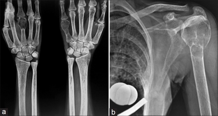
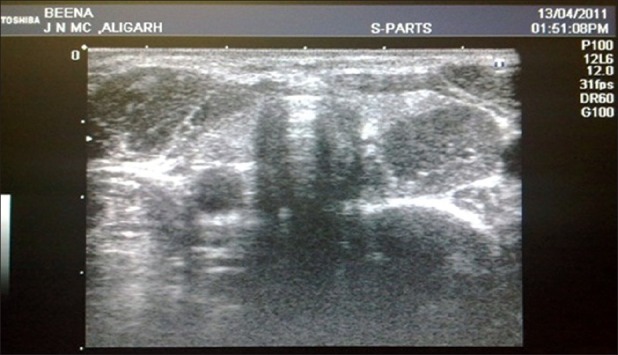
Primary hyperparathyroidism is a disease characterized by excessive secretion of parathormone. During the course of this disease, bone loss occurs, particularly depending on resorption of the skeletal system. One of the complications of primary hyperparathyroidism is fibrotic, cystic bony changes which is called Brown tumor. Skeletal manifestations in the form of Brown tumors are rare and according to literature occur in less than 2% of patients suffering from any form of hyperparathyroidism. Such rare and multiple benign lesions may simulate a malignant neoplasm and pose a real challenge for the clinician in its differential diagnosis. We present a case of a 23-year-old Indian woman who was evaluated for multiple lytic expansile lesions with a strong suspicion of malignancy and fibrous dysplasia but turned out to be a case of primary hyperparathyroidism.
Keywords: Brown tumors; endocrine system; expansile lesions; primary hyperparathyroidism.
Conflict of interest statement
Figures

References
-
- Van Herden JA, Beahrs OH, Woolner LB. The pathology and surgical management of primary Hyperparathyroidism. Surg Clin North Am. 1977;57:557–63. - PubMed
-
- Hsu CH, Liew PL, Wang W, Leung TK, Yang KM. Enhanced FDG uptake in brown tumors mimics multiple skeletal metastases in a patient with primary hyperparathyroidism. Acta Radiol. 2008;49:949–50. - PubMed
-
- Jouan A, Zabraniecki L, Vincent V, Poix E, Fournié B. An unusual presentation of primary Hyperparathyroidism: Severe hypercalcemia and multiple brown tumors. Joint Bone Spine. 2008;75:209–11. - PubMed
-
- Joyce JM, Idea RJ, Grossman SJ, Liss RG, Lyons JB. Multiple brown tumors in hyperparathyroidism mimicking metastatic disease on radiograph and bone scan. Clin Nucl Med. 1994;19:630–5. - PubMed
-
- Kalambokis G, Economou G, Kamina S, Papachristou DJ, Bai M, Tsianos EV. Multiple brown tumors of the ribs simulating malingnancy. J Endocrinol Invest. 2005;28:738–40. - PubMed