. 2012 Dec 5:6:82.
doi: 10.3389/fncir.2012.00082. eCollection 2012.

Neuronal nitric oxide synthase expressing neurons: a journey from birth to neuronal circuits

Affiliations

Neuronal nitric oxide synthase expressing neurons: a journey from birth to neuronal circuits

Ludovic Tricoire et al. Front Neural Circuits. .

Abstract

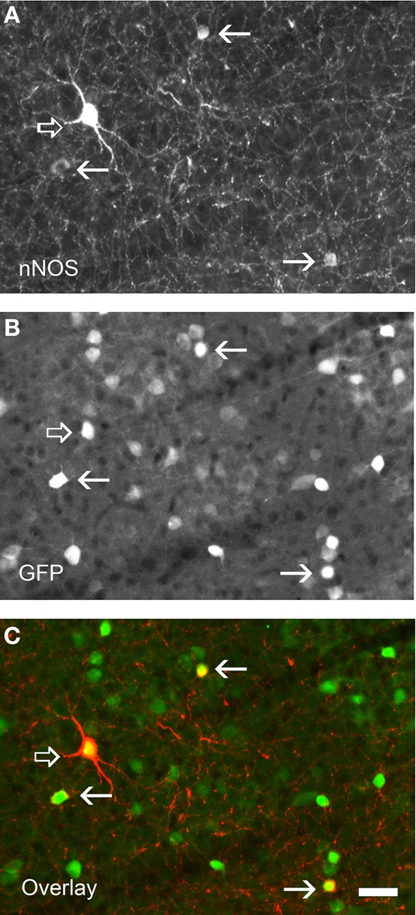
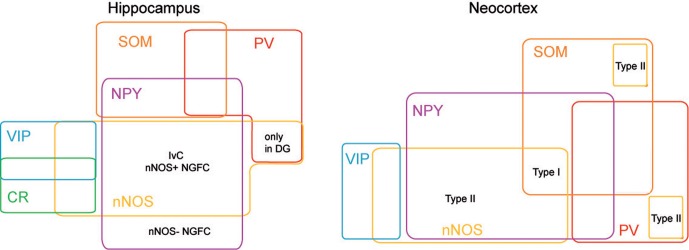
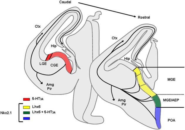
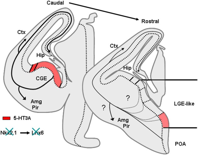
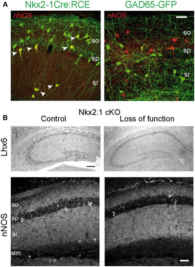
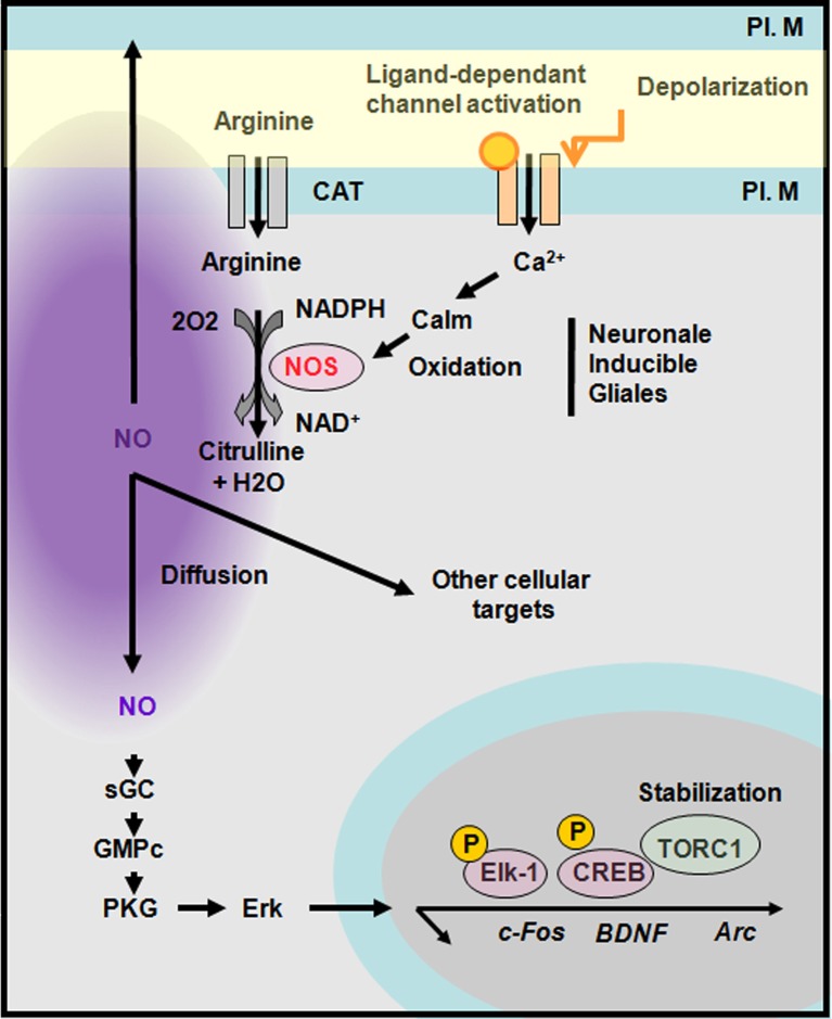
Nitric oxide (NO) is an important signaling molecule crucial for many physiological processes such as synaptic plasticity, vasomotricity, and inflammation. Neuronal nitric oxide synthase (nNOS) is the enzyme responsible for the synthesis of NO by neurons. In the juvenile and mature hippocampus and neocortex nNOS is primarily expressed by subpopulations of GABAergic interneurons. Over the past two decades, many advances have been achieved in the characterization of neocortical and hippocampal nNOS expressing neurons. In this review, we summarize past and present studies that have characterized the electrophysiological, morphological, molecular, and synaptic properties of these neurons. We also discuss recent studies that have shed light on the developmental origins and specification of GABAergic neurons with specific attention to neocortical and hippocampal nNOS expressing GABAergic neurons. Finally, we summarize the roles of NO and nNOS-expressing inhibitory neurons.

Keywords: GABA; classification; development; interneurons; nNOS; specification.


Figures

Figure 1
Figure 1
Examples of IvC, NGFC, and VIP+/nNOS+ interneurons. (A) Neurolucida reconstructions of biocytin-filled cells (black, dendrite; red, axon). (B) Voltage responses of cells shown in (A) to three current step injections (-200 pA, just suprathreshold, and twice the current for just suprathreshold). Adapted from Tricoire et al. (2010).
Figure 2
Figure 2
Immunolabeling for nNOS in a neocortical sections of GAD67:GFP mouse strain showing the two nNOS populations. (A) Fluorescence picture showing immunohistochemical expression of nNOS. (B) Expression pattern of GFP. (C) Overlay of (A) and (B). nNOS-type I neurons display strong immunolabeling (open arrows) and a large soma whereas nNOS-type II (arrows) are weakly stained and display smaller soma. Note that all nNOS-positive neurons are GABAergic. Scale bar: 30 μm. Unpublished caption obtained from preparations used for the study presented by Perrenoud et al. (2012a).
Figure 3
Figure 3
nNOS expressing interneurons in cortex and hippocampus. Scheme summarizing the molecular profiles of neocortical and hippocampal nNOS+ interneurons. This diagram is based on previous report (Tricoire et al., 2010, 2011) and on Perrenoud et al. ( in this issue).
Figure 4
Figure 4
Origins of GABAergic neurons populating the cerebral cortex and hippocampus at embryonic stages. Transversal schematic sections of E13–E14 embryonic mouse brain showing regions relevant to origin/birth of cortical interneurons. Territories expressing specific transcription factors or molecules classically used to determine the place of genesis of specific interneurons subpopulation are drawn. AEP, entopeduncular region, Amg, amygdala, CGE, caudal ganglionic eminence; Ctx, cortex; Hip, hippocampus; LGE, lateral ganglionic eminence; MGE, medial ganglionic eminence; Pir, piriform cortex; POA, preoptic area.
Figure 5
Figure 5
Phenotype of mice lacking Nkx2.1. Nkx2.1 knockout mice show a MGE respecified into a “LGE-like” territory. Since Nkx2.1 is necessary for Lhx6 expression, Lhx6 is not observed in these animals that lack most PV and SOM expressing neurons in the cortex and hippocampus. AEP, entopeduncular region, Amg, amygdala, CGE, caudal ganglionic eminence; Ctx, cortex; Hip, hippocampus; LGE, lateral ganglionic eminence; MGE, medial ganglionic eminence; Pir, piriform cortex; POA, preoptic area.
Figure 6
Figure 6
Embryonic origin of nNOS+ hippocampal interneurons. (A) Images illustrating the coexpression of GFP and nNOS in the Nkx2.1Cre:RCE (left) and GAD65-GFP (right) mouse lines. Scale bar: 25 μm. (B) Nkx2.1 is necessary for the specification of nNOS+ interneurons. Top, In situ hybridization against Lhx6 transcripts on hippocampus of control (left) and mutant (right) P15 mice after conditional loss of Nkx2.1 function at E10.5. Scale bar: 200 μm. Bottom, Immunohistochemical expression patterns of nNOS in CA1 of control and mutant mice. Scale bar: 50 μm. Adapted from Tricoire et al. (2010, 2011).
Figure 7
Figure 7
Synthesis of nitric oxide and transduction cascades. Neuronal nitric oxide synthase (nNOS) is activated by a calcium-dependant calmodulin. NOS produces nitric oxide (NO) upon oxidation of arginine into citrulline. NO diffuses and act on presynaptic or postsynaptic targets. A well-known pathway of NO is through the activation of guanylyl cyclase (GC) that activates a protein kinase G (PKG) leading to Erk activation and the stabilization of TORC1 a CREB co-activator. CAT, cation and anion transporter; PL. M, plasma membrane. Adapted from Gallo and Iadecola (2011).

Similar articles

Cited by

References

    1. Acsády L., Arabadzisz D., Freund T. F. (1996a). Correlated morphological and neurochemical features identify different subsets of vasoactive intestinal polypeptide-immunoreactive interneurons in rat hippocampus. Neuroscience 73, 299–315 10.1016/0306-4522(95)00610-9 - DOI - PubMed
    1. Acsády L., Görcs T. J., Freund T. F. (1996b). Different populations of vasoactive intestinal polypeptide-immunoreactive interneurons are specialized to control pyramidal cells or interneurons in the hippocampus. Neuroscience 173, 317–334 10.1016/0306-4522(95)00609-5 - DOI - PubMed
    1. Alcántara S., Ruiz M., D'Arcangelo G., Ezan F., de Lecea L., Curran T., et al. (1999). Regional and cellular patterns of reelin mRNA expression in the forebrain of the developing and adult mouse. J. Neurosci. 18, 7779–7799 - PMC - PubMed
    1. Armstrong C., Szabadics J., Tamás G., Soltesz I. (2011). Neurogliaform cells in the molecular layer of the dentate gyrus as feed-forward γ-aminobutyric acidergic modulators of entorhinal-hippocampal interplay. J. Comp. Neurol. 519, 1476–1491 10.1002/cne.22577 - DOI - PMC - PubMed
    1. Arnold W. P., Mittal C. K., Katsuki S., Murad F. (1997). Nitric oxide activates guanylate cyclase and increases guanosine 3′:5′-cyclic monophosphate levels in various tissue preparations. Proc. Natl. Acad. Sci. U.S.A. 74, 3203–3207 - PMC - PubMed