Endoscopic ultrasound-guided fine needle aspiration of the celiac ganglion: A diagnostic pitfall
- PMID: 23227103
- PMCID: PMC3513782
- DOI: 10.4103/1742-6413.103025
Endoscopic ultrasound-guided fine needle aspiration of the celiac ganglion: A diagnostic pitfall
Abstract
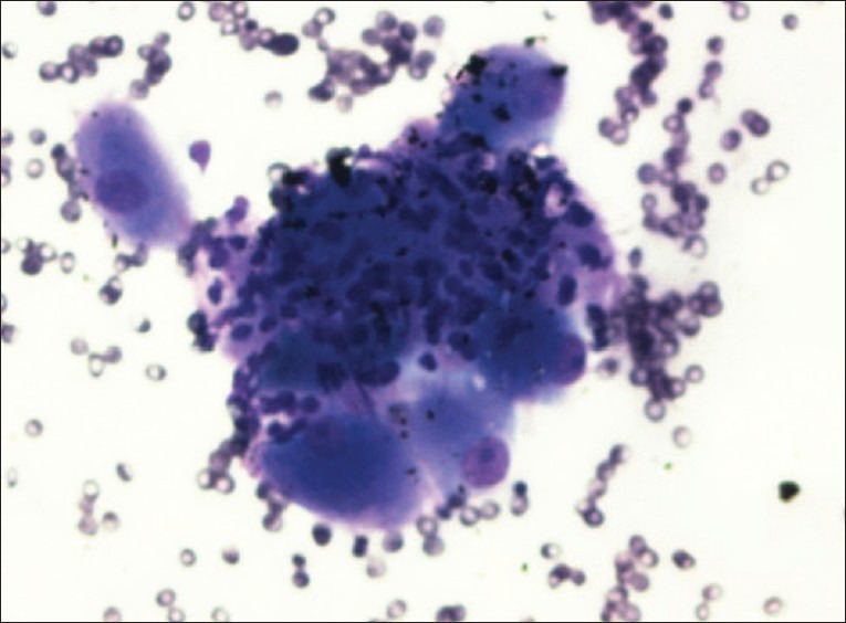
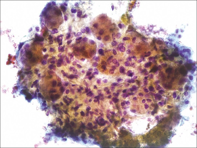
Endoscopic ultrasound guided fine-needle aspiration (EUS-FNA) is now widely used as a primary tool in the evaluation of lymphadenopathy in both the mediastinum and abdomen. A sympathetic ganglion may be mistaken for an enlarged lymph node on endoscopic ultrasound and are rarely sampled as such. A 51-year-old female presented with a history of weight loss, vomiting for several months, and right upper quadrant discomfort. Computed tomography (CT) and magnetic resonance imaging (MRI) scans showed a dilated common bile duct (CBD) with a possible periampullary mass, paraaortic, and pericelial lymph nodes suspicious for metastatic disease. Endosonography revealed a 17 mm oval hypoechoic structure with distinct margins in the para-aortic, celiac axis region suggestive of an enlarged lymph node. An EUS-FNA was done. Cytology revealed ganglion cells with large oval epithelial-like cells with round nuclei and prominent nucleoli consistent with a benign sympathetic ganglion. It is crucial for the cytopathologist to be aware of the fact that the endoscopist might have sampled a celiac ganglion instead of a celiac lymph node and be able to distinguish the cytological features of a benign sympathetic ganglion from a malignant process.
Keywords: Celiac ganglion; endoscopic ultrasound; fine needle aspiration.
Figures
Similar articles
-
Frequency and characterization of celiac ganglia diagnosed on fine-needle aspiration.Cytojournal. 2015 Feb 18;12:4. doi: 10.4103/1742-6413.151677. eCollection 2015. Cytojournal. 2015. PMID: 25745503 Free PMC article.
-
A comparison of the accuracy of echo features during endoscopic ultrasound (EUS) and EUS-guided fine-needle aspiration for diagnosis of malignant lymph node invasion.Gastrointest Endosc. 1997 Jun;45(6):474-9. doi: 10.1016/s0016-5107(97)70176-7. Gastrointest Endosc. 1997. PMID: 9199903
-
Primary Amyloidosis of Celiac/Para-Pancreatic Lymph Nodes Diagnosed by Endosonography-Guided Fine Needle Aspiration: A Case Report.J Investig Med High Impact Case Rep. 2015 Sep 24;3(3):2324709615607916. doi: 10.1177/2324709615607916. eCollection 2015 Jul-Sep. J Investig Med High Impact Case Rep. 2015. PMID: 26904706 Free PMC article.
-
Needle tract seeding recurrence of pancreatic cancer in the gastric wall with paragastric lymph node metastasis after endoscopic ultrasound-guided fine needle aspiration followed by pancreatectomy: a case report and literature review.BMC Gastroenterol. 2020 Jan 15;20(1):13. doi: 10.1186/s12876-020-1159-x. BMC Gastroenterol. 2020. PMID: 31941458 Free PMC article. Review.
-
Endoscopic ultrasound fine needle aspiration vs fine needle biopsy for pancreatic masses, subepithelial lesions, and lymph nodes.World J Gastroenterol. 2021 Jul 14;27(26):4194-4207. doi: 10.3748/wjg.v27.i26.4194. World J Gastroenterol. 2021. PMID: 34326619 Free PMC article. Review.
Cited by
-
Announcement of first time Cytojournal impact factor for 2012 coincides with Cytojournal decade celebration (2004-2013).Cytojournal. 2013 Aug 30;10:18. doi: 10.4103/1742-6413.117359. eCollection 2013. Cytojournal. 2013. PMID: 24082914 Free PMC article. No abstract available.
-
Frequency and characterization of celiac ganglia diagnosed on fine-needle aspiration.Cytojournal. 2015 Feb 18;12:4. doi: 10.4103/1742-6413.151677. eCollection 2015. Cytojournal. 2015. PMID: 25745503 Free PMC article.
References
-
- Wang KX, Ben QW, Jin ZD, Du YQ, Zou DW, Liao Z, et al. Assessment of morbidity and mortality associated with EUS-guided FNA: A systematic review. Gastrointest Endosc. 2011;73:283–90. - PubMed
-
- Galasso D, Carnuccio A, Larghi A. Pancreatic cancer: Diagnosis and endoscopic staging. Eur Rev Med Pharmacol Sci. 2010;14:375–85. - PubMed
-
- Jamil LH, Gill KR, Wallace MB. Staging and restaging of advanced esophageal cancer. Curr Opin Gastroenterol. 2008;24:530–4. - PubMed
-
- Gill KR, Wallace MB. Endoscopic ultrasound and staging of non-small cell lung cancer. Minerva Med. 2007;98:323–30. - PubMed
-
- Knight CS, Eloubeidi MA, Crowe R, Jhala NC, Jhala DN, Chhieng DC, et al. Utility of endoscopic ultrasound-guided fine-needle aspiration in the diagnosis and staging of colorectal carcinoma. Diagn Cytopathol. 2011 Sep 19; doi: 10.1002/dc.21804. [PMID: 21932358] - PubMed
Publication types
LinkOut - more resources
Full Text Sources