Review
. 2012 Apr;2(2):1541-84.
doi: 10.1002/cphy.c110052.

Regulation of transport in the connecting tubule and cortical collecting duct

Affiliations
Review

Regulation of transport in the connecting tubule and cortical collecting duct

Alexander Staruschenko. Compr Physiol. 2012 Apr.

Abstract

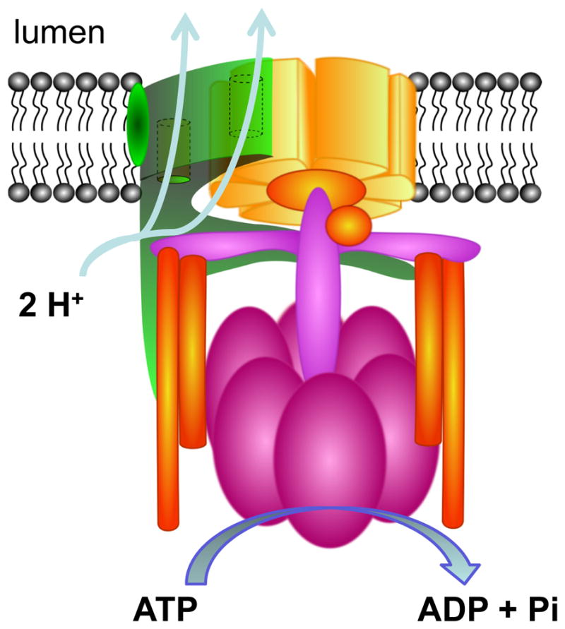
The central goal of this overview article is to summarize recent findings in renal epithelial transport,focusing chiefly on the connecting tubule (CNT) and the cortical collecting duct (CCD).Mammalian CCD and CNT are involved in fine-tuning of electrolyte and fluid balance through reabsorption and secretion. Specific transporters and channels mediate vectorial movements of water and solutes in these segments. Although only a small percent of the glomerular filtrate reaches the CNT and CCD, these segments are critical for water and electrolyte homeostasis since several hormones, for example, aldosterone and arginine vasopressin, exert their main effects in these nephron sites. Importantly, hormones regulate the function of the entire nephron and kidney by affecting channels and transporters in the CNT and CCD. Knowledge about the physiological and pathophysiological regulation of transport in the CNT and CCD and particular roles of specific channels/transporters has increased tremendously over the last two decades.Recent studies shed new light on several key questions concerning the regulation of renal transport.Precise distribution patterns of transport proteins in the CCD and CNT will be reviewed, and their physiological roles and mechanisms mediating ion transport in these segments will also be covered. Special emphasis will be given to pathophysiological conditions appearing as a result of abnormalities in renal transport in the CNT and CCD.


Figures

Figure 1
Figure 1
Consecutive segments of the nephron. Tubules discussed in this overview article are highlighted in red.
Figure 2
Figure 2
(A) Structure of the nephron. Abbreviations for the nephron segments are described in Fig. 1. The relative lengths of different segments are not drawn to scale. (B) The ICT and CCD are composed of principal and intercalated cells. Structure of the CCD shown as a cross-section and schematic presentation of principal and intercalated cells that comprise these segments. The connecting tubule cells and the principal cells have a polygonal shape. The intercalated cells have a rounded shape. Compared to intercalated cells, the connecting tubule cells and the principal cells have fewer mitochondria and only modestly develop invaginations of the basolateral membrane. Both types of cells develop apical microvilli. However, primary cilia is found only in principal cells.
Figure 3
Figure 3
Representative immunohistochemical staining for AQP2 in the cortical sections of the Sprague-Dawley rat kidney. Original magnifications are 20x and 60x, scale bars are shown on the pictures. Negative controls (stained with secondary antibodies in the absence of primary antibodies or stained without primary and secondary antibodies) did not have any staining (data not shown). Several profiles of proximal tubules (PT), distal convoluted tubules (DCT), cortical collecting duct (CCD) and glomerulus (G) are marked by arrows at 20x. Representative examples of intercalated (IC; no staining) and principal (PC; stained for AQP2, shown in brown) cells are indicated on the close-up image. The kidney was fixed for 24 hrs in zinc formalin and processed for paraffin embedding as described previously (240, 314, 413). The kidney sections were cut, dried and deparaffinized for subsequent labeled streptavidin-biotin immunohistochemistry. All slides were counterstained with Mayer’s hematoxylin (Dako, Carpinteria, CA). Tissue sections were incubated for 45 min in a 1:200 concentration of anti-AQP2 (sc-28629; Santa Cruz Biotechnology).
Figure 4
Figure 4
Primary transport characteristics of the cortical collecting duct. Principal and intercalated cells are in colored beige and green, respectively. Tight junctions are also schematically represented in between the cells.
Figure 5
Figure 5
Major channels and transporters involved in water and electrolyte homeostasis in principal cells of the CCD.
Figure 6
Figure 6
Mechanism of action of aldosterone in principal cells of the CCD. Aldosterone (Aldo) binds to mineralocorticoid receptor (MR) that then translocates to the nucleus and upregulates transcription of aldosterone-induced proteins, which regulate sodium reabsorption and potassium secretion via affecting ENaC, ROMK and Na+/K+-ATPase. Effect of 11β-HSD2 that metabolizes cortisol to cortisone, which has little affinity for MR or glucocorticoid receptor (GR), is shown.
Figure 7
Figure 7
Predicted structure for human ENaC (hENaC) based on the structure for cASIC1 (248). (A) Predicted subunit structure for α-subunit of hENaC. Predicted domain organization of adjusted α-hENaC modeled on the cASIC1 A monomer (using 2QTS coordinates). Secondary structure, domain labeling and coloring follows that used by Jasti and colleagues for cASIC1 (248): transmembrane domains TM1 and TM2 and linker regions red, palm is yellow, β-ball orange, knuckle cyan, and thumb green. The exception is that the finger domain is magenta and blue. Blue highlights areas of hENaC that likely have marked differences compared to cASIC1. Putative disulfide bridges are labeled 1–7 and shown as yellow sticks. The conserved Trp87 (green side chain) at the beginning of TM1 and Tyr391 (red side chain) within the putative coupling loop are shown. Conserved Ser115 and Glu538 possibly involved in intrasubunit H-bond formation are shown with red side chains. (B) View of the ribbon structure of the predicted heterotrimeric hENaC. Adapted α- (red), β- (yellow), and γ-(blue) hENaC modeled using the 2QTS structural coordinates for the A, B, and C subunits of the cASIC1 homotrimer. Figure is adapted from (542) with permission.
Figure 8
Figure 8
Structure and distribution of ROMK channels. (A) Schematic presentation of ROMK structure shows two characteristic transmembrane segments (TM1 and TM2; blue and green, respectively), NH2- and COOH-termini and an extracellular domain. (B) Predicted structure of ROMK subunit stoichiometry. (C) Distribution of ROMK isoforms expression along the nephron. TAL, thick ascending limb of Henle’s loop; DCT, distal convoluted tubule; CNT, connecting tubule; CCD, cortical collecting duct; OMCD, outer medullary collecting duct.
Figure 9
Figure 9
The structure of the pore-forming α-subunit and regulatory β-subunit of the BK channel (A). α-subunit contains 7 putative transmembrane domains, S0–S6, a conserved K+-selective pore region between S5 and S6, and a long COOH-terminal cytosolic tail. β-subunit contains two transmembrane segments and short NH2- and COOH-termini. (B) Proposed model of BK channel. Four BK α-subunits co-assemble with four BK β-subunits to form the channel heteromultimer.
Figure 10
Figure 10
Types of intercalated cells. Type A intercalated cells secrete H+ via a V-type H+-ATPase in the apical plasma membrane and transport HCO3 in exchange for Cl via basolateral Cl/HCO3 exchangers including the AE1 anion exchanger. Type B intercalated cells exhibit an inverse functional polarity to that of type A intercalated cells. A non-A, non-B type co-express Cl/HCO3 exchangers such as pendrin and H+-ATPase in the apical plasma membrane. Schematic representations of the V-ATPse, pendrin and AE-2 are shown in Figs. 11 and 12.
Figure 11
Figure 11
Scheme of the V-type H+-ATPase. H+-ATPases use the energy released by the hydrolysis of ATP to move protons against their concentration gradients. The V0 domain is involved in translocation of the protein. The V1 domain is involved in ATP-hydrolysis. The precise subunits composition is not entirely clear and several slightly different schemes are proposed. For details see recent excellent reviews providing details about subunits and domains of V-type H+-ATPase (68, 162, 474, 635).
Figure 12
Figure 12
Proposed schematics of Cl/HCO3 exchangers. The structures of anion exchanger AE1 (SLC4A1) (A) and pendrin (SLC26A4) (B) are shown. As seen from these schemes, both NH2- and COOH-termini of AE1 are intracellular. In contrast, COOH-terminus of pendrin is extracellular.
Figure 13
Figure 13
Regulation of the aquaporin-2 (AQP2)-mediated water transport by arginine vasopressin (AVP). (A) Proposed topology of AQP2. An AQP2 monomer consists of six transmembrane domains connected by five loops. NH2- and COOH-termini are located intracellularly. (B) A scheme of water transport regulation by AVP. Vasopressin receptor (V2R), stimulatory GTP-binding protein (Gs), adenylate cyclase (AC), adenosine triphosphate (ATP), and cyclic adenosine monophosphate (cAMP) are indicated.
Figure 14
Figure 14
Transmembrane topology of TRP channels. TRP channels belong to the large superfamily of cation channels with six transmembrane-spanning segments forming a transmembrane domain with a pore loop inserted between TM5 and TM6 and NH2- and COOH-intracellular termini (A). In contrast, polycystin 1 (PKD1 or PC1) has eleven transmembrane domains and a large extracellular NH2 domain (B).
Figure 15
Figure 15
Mutations in genes encoding proteins functionally expressed in CCD cause severe kidney disorders. (A) Mechanisms of Cyst Formation in autosomal dominant (ADPKD) and recessive (ARPKD) polycystic kidney diseases. Normal tubule is also shown. Representative images of kidney cortical sections of Sprague-Dawley (B) and PCK (C) rats. PCK rat, a model of ARPKD, demonstrates abundant formation of cysts. Original magnifications are 40x. Scale bar is presented.
Figure 16
Figure 16
Modes of transepithelial transport and major proteins involved in the paracellular transport. Schemes of the transcellular (A) and paracellular (B) epithelial transport. (C) Schematic three-dimensional structure of tight junctions. Proposed structures of claudin (D) and occludin (E).

References

    1. Abe K, Tani K, Fujiyoshi Y. Conformational rearrangement of gastric H+,K+-ATPase induced by an acid suppressant. Nat Commun. 2011;2:155. - PMC - PubMed
    1. Abe K, Tani K, Nishizawa T, Fujiyoshi Y. Inter-subunit interaction of gastric H+,K+-ATPase prevents reverse reaction of the transport cycle. EMBO J. 2009;28:1637–1643. - PMC - PubMed
    1. Abramowitz J, Birnbaumer L. Physiology and pathophysiology of canonical transient receptor potential channels. FASEB J. 2009;23:297–328. - PMC - PubMed
    1. Adachi S, Uchida S, Ito H, Hata M, Hiroe M, Marumo F, Sasaki S. Two isoforms of a chloride channel predominantly expressed in thick ascending limb of Henle’s loop and collecting ducts of rat kidney. J Biol Chem. 1994;269:17677–17683. - PubMed
    1. Ahn KY, Turner PB, Madsen KM, Kone BC. Effects of chronic hypokalemia on renal expression of the “gastric” H+,K+-ATPase alpha-subunit gene. Am J Physiol Renal Physiol. 1996;270:F557–F566. - PubMed

Publication types

MeSH terms

LinkOut - more resources