. 2012:2012:823534.
doi: 10.1155/2012/823534. Epub 2012 Nov 25.

H3K79me3T80ph is a Novel Histone Dual Modification and a Mitotic Indicator in Melanoma

Affiliations

H3K79me3T80ph is a Novel Histone Dual Modification and a Mitotic Indicator in Melanoma

Danielle R Martinez et al. J Skin Cancer. 2012.

Abstract

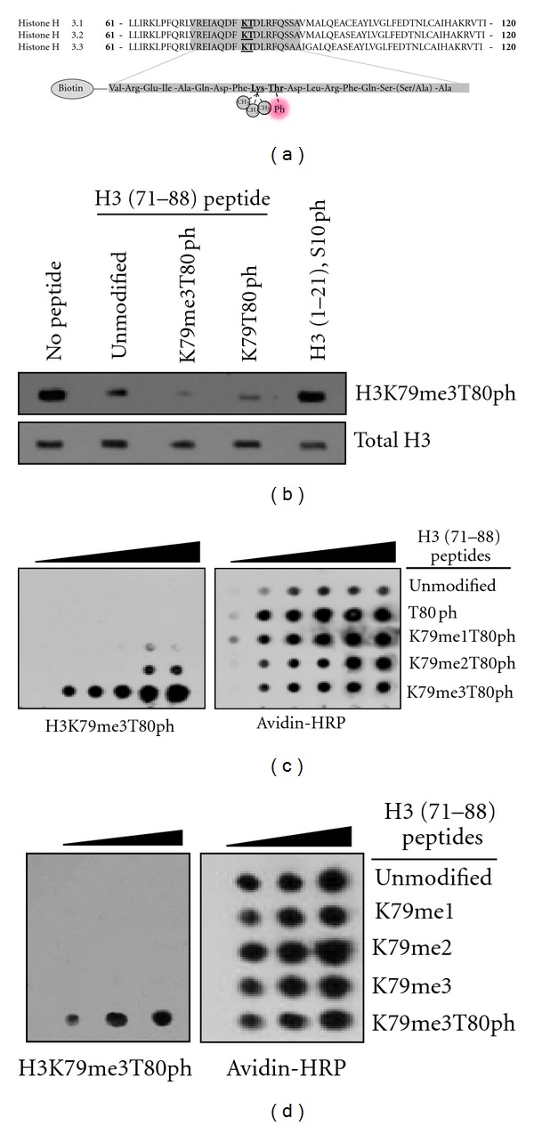
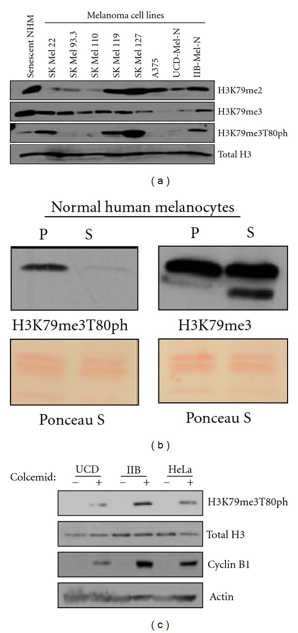
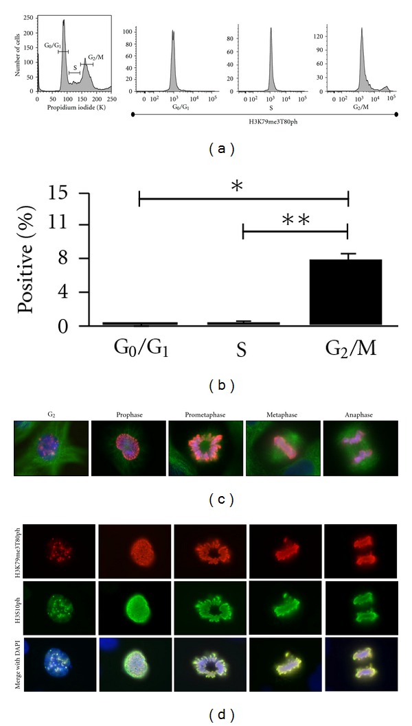
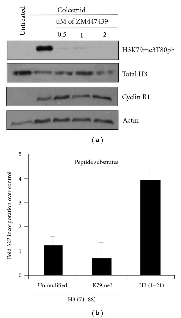
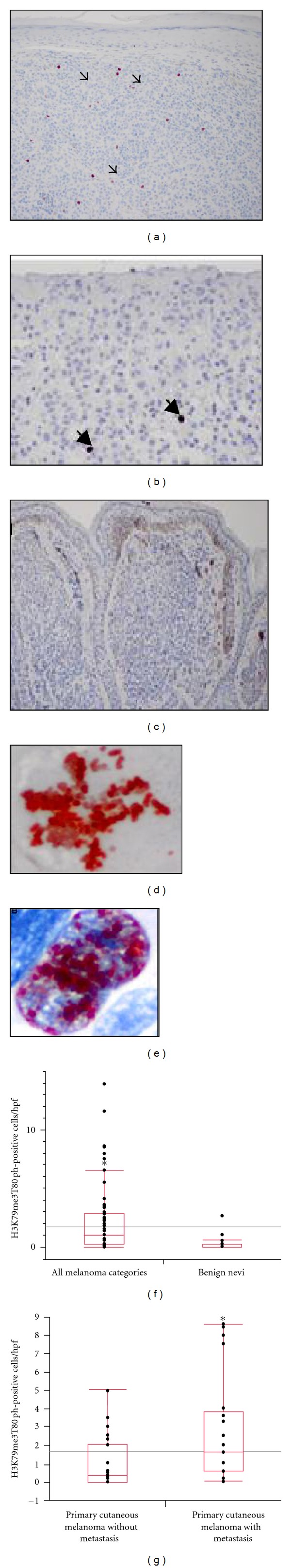
The current study characterizes the mitosis-associated histone dual modification on the core of histone H3: trimethylation of histone H3 lysine 79 and simultaneous phosphorylation of H3 threonine 80 (H3K79me3T80ph). Through the use of protein and microscopy-based techniques, we find that H3K79me3T80ph shares a similar spatial and temporal regulation as H3S10ph but additionally requires methyltransferase activity. In addition, we find that Aurora kinase activity is necessary for the catalysis of H3K79me3T80ph in vivo. Finally, our analysis of H3K79me3T80ph using a tissue microarray indicates that H3K79me3T80ph marks a subset of primary cutaneous melanomas with metastatic potential indicating that H3K79me3T80ph may identify a subset of invasive melanomas with a more aggressive clinical behaviour.


Figures

Figure 1
Figure 1
H3K79me3T80ph antibody specificity. (a) Shown are the three H3 isoforms and their amino acid sequence in the core of the protein surrounding lysine 79 and threonine 80 (emphasized) (top), and the biotinylated peptides designed for this study that are either unmodified, contain mono-, di-, or trimethylation of K79 plus phosphorylated T80, or phosphorylated T80 alone (bottom). (b) Histones from HeLa cells were isolated and subjected to western blotting with the H3K79me3T80ph antibody alone (no peptide) or in the presence of 15 µM of biotinylated H3 (71–88) peptides with no modification, K79 trimethylation and T80 phosphorylation, T80 phosphorylation alone, or an H3 (1–21) S10ph peptide. (c) 50, 100, 150, 200, 250 nmoles of biotinylated H3 (71–88) peptides containing mono-, di-, or trimethylated K79 in conjunction with T80 phosphorylation were spotted onto nitrocellulose and subjected to H3K79me3T80ph and Avidin-HRP western blotting. (d) 50, 100, 150, 200, 250 nmoles of biotinylated H3 (71–88) peptides with K79 trimethylation and T80 phosphorylation or mono-, di-, or trimethylated K79 alone were spotted onto nitrocellulose and subjected to H3K79me3T80ph and avidin-HRP western blotting.
Figure 2
Figure 2
H3K79me3T80ph is cell cycle regulated. (a) H3K79me2, H3K79me3, and H3K79me3T80ph western blots of histones isolated from nondividing senescent human melanocytes and various human melanoma cell lines. (b) Western blot comparison of H3K79me3 and H3K79me3T80ph levels on histones from proliferating (P) and senescent (S) normal human melanocytes. (c) UCD-Mel-N, IIB-Mel-N, and HeLa cells were either asynchronous (−) or mitotically synchronized using colcemid (+). Histones were isolated to perform an H3K79me3T80ph western blot, and whole cell lysate was used to perform Cyclin B1 western blotting as a control for cell synchronization.
Figure 3
Figure 3
H3K79me3T80ph is mitosis-associated. (a) Representative flow cytometry experiment of untreated asynchronous HeLa cells stained with propidium iodide and H3K79me3T80ph (Area). Propidium iodide incorporation was used to obtain a cell cycle profile, and the cell cycle phases were then gated. An H3K79me3T80ph histogram was created for each phase of the cell cycle. (b) Percentage of H3K79me3T80ph positive cells in each cell cycle phase as determined by flow cytometry of asynchronous HeLa cells stained with propidium iodide and H3K79me3T80ph (Area). Bars represent mean values (± S.E.M.), n = 3; *p G0/G1−G2/M = 0.0014, **p S−G2/M = 0.0016. (C) Immunofluorescence of asynchronous UCD-Mel-N cells stained for H3K79me3T80ph (red), tubulin (green), and DNA (blue). (d) H3K79me3T80ph and H3S10ph co-immunofluorescence performed on asynchronous UCD-Mel-N cells; H3K79me3T80ph (red), H3S10ph (green), DNA (blue).
Figure 4
Figure 4
Aurora B is required for H3T80 phosphorylation. (a) HeLa cells were subjected to no treatment, colcemid arrest, and colcemid arrest in conjunction with ZM447439 treatment. Half of the cells were used to isolate histones and to perform an H3K79me3T80ph western blot, and half of the cells were used to make whole-cell lysate and perform a Cyclin B1 western to control for synchronization. (b) In vitro kinase assay using recombinant Aurora B/INCENP in the presence of 32P-gamma-ATP and biotinylated H3 (1–21); H3 (71–88); H3 (71–88), K79me3 peptides. Bars represent fold mean 32P scintillation counts over cold phosphorylated peptide (±S.E.M.), n = 3.
Figure 5
Figure 5
Immunohistochemistry (IHC) H3K79me3T80ph detects a subset of primary cutaneous melanomas with metastatic potential. Positive H3K79me3T80ph cells (arrows) in: (a) metastatic melanoma of the skin (magnification: ×100) and (b) primary cutaneous invasive melanoma (magnification: ×200) (c) Melanocytic nevi with absence of H3K79me3T80ph labeling (magnification: ×100). (d) and (e) Representative H3K79me3T80ph staining of melanoma tissue microarray (d): mitotically active cells, (e): melanoma cells with speckled nuclear staining pattern (Magnification: ×400). (f) and (g) Box graph analysis of the number of H3K79me3T80ph positive cells per high powered field (h.p.f.) within the melanocytic neoplasms contained in the tissue microarray. (f) Melanoma from all categories demonstrates greater number H3K79me3T80ph positive cells compared to benign nevi (P = 0.0019). (g) Significant labelling of H3K79me3T80ph positive cells in primary cutaneous melanomas with known metastasis compared to primary melanomas without metastasis (P = 0.0044).

References

    1. Zhang Y, Reinberg D. Transcription regulation by histone methylation: interplay between different covalent modifications of the core histone tails. Genes and Development. 2001;15(18):2343–2360. - PubMed
    1. Feng Q, Wang H, Ng HH, et al. Methylation of H3-lysine 79 is mediated by a new family of HMTases without a SET domain. Current Biology. 2002;12(12):1052–1058. - PubMed
    1. Spellman PT, Sherlock G, Zhang MQ, et al. Comprehensive identification of cell cycle-regulated genes of the yeast Saccharomyces cerevisiae by microarray hybridization. Molecular Biology of the Cell. 1998;9(12):3273–3297. - PMC - PubMed
    1. Schulze JM, Jackson J, Nakanishi S, et al. Linking cell cycle to histone modifications: SBF and H2B monoubiquitination machinery and cell-cycle regulation of H3K79 dimethylation. Molecular Cell. 2009;35(5):626–641. - PMC - PubMed
    1. Jones B, Su H, Bhat A, et al. The histone H3K79 methyltransferase Dot1L is essential for mammalian development and heterochromatin structure. PLoS Genetics. 2008;4(9)e1000190 - PMC - PubMed

LinkOut - more resources