

An induction of microRNA, miR-7 through estrogen treatment in breast carcinoma
- PMID: 23227519
- PMCID: PMC3445861
- DOI: 10.1186/1479-5876-10-s1-s2
An induction of microRNA, miR-7 through estrogen treatment in breast carcinoma
Abstract
Background: Estrogen plays an important role in the development of estrogen-dependent breast carcinoma. Recently, several studies demonstrated a possible involvement of several micro RNAs (miRNAs) in the development of resistance to endocrine therapy in breast cancer patients, but the correlation between estrogen actions and miRNA expression in breast carcinoma still remains largely unknown. Therefore, in this study, we examined the in vitro effects of estrogen upon miRNA expression profiles in breast carcinoma.
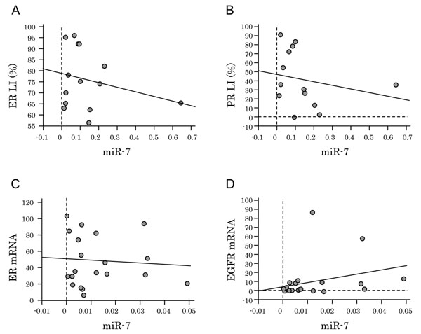
Methods: We first screened the miRNA expression profiles induced by 17β-Estradiol (E2) using RT2 miRNA PCR Array in the ER-positive breast carcinoma cell line MCF-7. We identified miR-7 as the important miRNA associated with estrogen actions in these cells and further examined the changes of estrogen-dependent EGFR expression by miR-7 in ER-positive or -negative breast carcinoma cell lines including MCF-7. We also evaluated the correlation between miR-7 and EGFR expression in breast carcinoma cells derived from 21 patients using laser capture microdissection combined with quantitative reverse transcriptase-PCR.
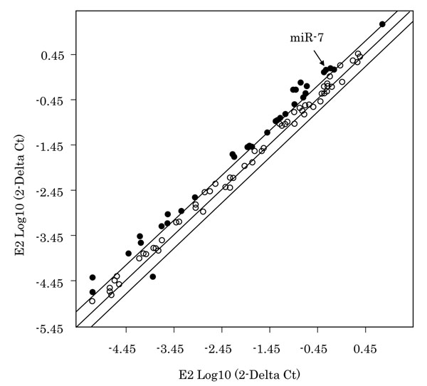
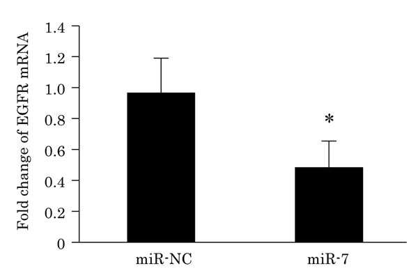
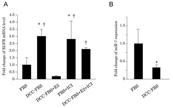
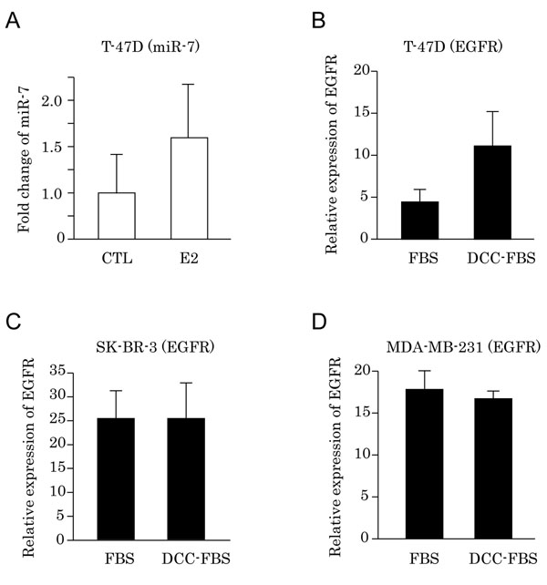
Results: Seventeen miRNAs were significantly induced by E2 treatment in the MCF-7 cell line. Among 17 miRNAs induced by estradiol treatment, only miR-7 expression was significantly decreased by subsequent ICI treatment. The expression of miR-7 was up-regulated 2.94-fold by E2 treatment. miR-7 was reported to suppress epidermal growth factor receptor (EGFR) expression in several human malignancies. Transfection of miR-7 significantly suppressed EGFR mRNA levels in MCF-7 cells. Depletion of E2 from cell culture media also increased the expression level of EGFR mRNA in MCF-7 and T-47D cells but not in ER-negative, MDA-MB-231 and SK-BR-3 cells. We also evaluated the status of miR-7 in breast carcinoma tissues, but the correlation between the status of miR-7 and EGFR in carcinoma cells isolated by laser capture microscopy was not detected.
Conclusions: These results suggest that miR-7 may play a role in the development of resistance to endocrine therapy in breast cancer patients through regulating EGFR expression of carcinoma cells.
Figures
Similar articles
-
Induction of cell proliferation and survival genes by estradiol-repressed microRNAs in breast cancer cells.BMC Cancer. 2012 Jan 20;12:29. doi: 10.1186/1471-2407-12-29. BMC Cancer. 2012. PMID: 22260523 Free PMC article.
-
Widespread estrogen-dependent repression of micrornas involved in breast tumor cell growth.Cancer Res. 2009 Nov 1;69(21):8332-40. doi: 10.1158/0008-5472.CAN-09-2206. Epub 2009 Oct 13. Cancer Res. 2009. PMID: 19826037 Clinical Trial.
-
Analysis of the miRNA-mRNA-lncRNA networks in ER+ and ER- breast cancer cell lines.J Cell Mol Med. 2015 Dec;19(12):2874-87. doi: 10.1111/jcmm.12681. Epub 2015 Sep 28. J Cell Mol Med. 2015. PMID: 26416600 Free PMC article.
-
miRNAs regulated by estrogens, tamoxifen, and endocrine disruptors and their downstream gene targets.Mol Cell Endocrinol. 2015 Dec 15;418 Pt 3(0 3):273-97. doi: 10.1016/j.mce.2015.01.035. Epub 2015 Feb 3. Mol Cell Endocrinol. 2015. PMID: 25659536 Free PMC article. Review.
-
The Divergent Effects of Ovarian Steroid Hormones in the MCF-7 Model for Luminal A Breast Cancer: Mechanistic Leads for Therapy.Int J Mol Sci. 2022 Apr 27;23(9):4800. doi: 10.3390/ijms23094800. Int J Mol Sci. 2022. PMID: 35563193 Free PMC article. Review.
Cited by
-
Serum miR-181d-5p levels in response to controlled ovarian stimulation: predictive value and biological function.JBRA Assist Reprod. 2023 Sep 12;27(3):391-400. doi: 10.5935/1518-0557.20220053. JBRA Assist Reprod. 2023. PMID: 36952624 Free PMC article.
-
Abnormal expression of miR-1 in breast carcinoma as a potent prognostic factor.Cancer Sci. 2015 Nov;106(11):1642-50. doi: 10.1111/cas.12808. Epub 2015 Oct 7. Cancer Sci. 2015. PMID: 26331797 Free PMC article.
-
Clinical Theragnostic Relationship between Drug-Resistance Specific miRNA Expressions, Chemotherapeutic Resistance, and Sensitivity in Breast Cancer: A Systematic Review and Meta-Analysis.Cells. 2019 Oct 14;8(10):1250. doi: 10.3390/cells8101250. Cells. 2019. PMID: 31615089 Free PMC article.
-
Molecular mechanism of the miR-7/BCL2L1/P53 signaling axis regulating the progression of hepatocellular carcinoma.Ann Transl Med. 2023 Jan 15;11(1):12. doi: 10.21037/atm-22-5929. Epub 2023 Jan 10. Ann Transl Med. 2023. PMID: 36760243 Free PMC article.
-
Effects of Yangzheng Sanjie Decoction-containing serum mediated by microRNA-7 on cell proliferation and apoptosis in gastric cancer.Oncol Lett. 2018 Mar;15(3):3621-3629. doi: 10.3892/ol.2018.7757. Epub 2018 Jan 9. Oncol Lett. 2018. PMID: 29467883 Free PMC article.
References
-
- Massarweh S, Schiff R. Resistance to endocrine therapy in breast cancer: exploiting estrogen receptor/growth factor signaling crosstalk. Endocr Relat Cancer. 2006;13(Suppl 1):S15–24. - PubMed
MeSH terms
Substances
LinkOut - more resources
Full Text Sources
Medical
Research Materials
Miscellaneous
