Bupivacaine-induced apoptosis independently of WDR35 expression in mouse neuroblastoma Neuro2a cells
- PMID: 23227925
- PMCID: PMC3541351
- DOI: 10.1186/1471-2202-13-149
Bupivacaine-induced apoptosis independently of WDR35 expression in mouse neuroblastoma Neuro2a cells
Abstract
Background: Bupivacaine-induced neurotoxicity has been shown to occur through apoptosis. Recently, bupivacaine was shown to elicit reactive oxygen species (ROS) production and induce apoptosis accompanied by activation of p38 mitogen-activated protein kinase (MAPK) in a human neuroblastoma cell line. We have reported that WDR35, a WD40-repeat protein, may mediate apoptosis through caspase-3 activation. The present study was undertaken to test whether bupivacaine induces apoptosis in mouse neuroblastoma Neuro2a cells and to determine whether ROS, p38 MAPK, and WDR35 are involved.
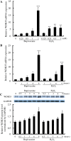
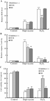
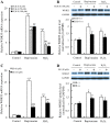
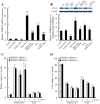
Results: Our results showed that bupivacaine induced ROS generation and p38 MAPK activation in Neuro2a cells, resulting in apoptosis. Bupivacaine also increased WDR35 expression in a dose- and time-dependent manner. Hydrogen peroxide (H(2)O(2)) also increased WDR35 expression in Neuro2a cells. Antioxidant (EUK-8) and p38 MAPK inhibitor (SB202190) treatment attenuated the increase in caspase-3 activity, cell death and WDR35 expression induced by bupivacaine or H(2)O(2). Although transfection of Neuro2a cells with WDR35 siRNA attenuated the bupivacaine- or H(2)O(2)-induced increase in expression of WDR35 mRNA and protein, in contrast to our previous studies, it did not inhibit the increase in caspase-3 activity in bupivacaine- or H(2)O(2)-treated cells.
Conclusions: In summary, our results indicated that bupivacaine induced apoptosis in Neuro2a cells. Bupivacaine induced ROS generation and p38 MAPK activation, resulting in an increase in WDR35 expression, in these cells. However, the increase in WDR35 expression may not be essential for the bupivacaine-induced apoptosis in Neuro2a cells. These results may suggest the existence of another mechanism of bupivacaine-induced apoptosis independent from WDR35 expression in Neuro2a cells.
Figures


Similar articles
-
Enhanced expression of WD repeat-containing protein 35 via nuclear factor-kappa B activation in bupivacaine-treated Neuro2a cells.PLoS One. 2014 Jan 21;9(1):e86336. doi: 10.1371/journal.pone.0086336. eCollection 2014. PLoS One. 2014. PMID: 24466034 Free PMC article.
-
Enhanced expression of WD repeat-containing protein 35 via CaMKK/AMPK activation in bupivacaine-treated Neuro2a cells.PLoS One. 2014 May 23;9(5):e98185. doi: 10.1371/journal.pone.0098185. eCollection 2014. PLoS One. 2014. PMID: 24859235 Free PMC article.
-
Bupivacaine induces apoptosis via mitochondria and p38 MAPK dependent pathways.Eur J Pharmacol. 2011 Apr 25;657(1-3):51-8. doi: 10.1016/j.ejphar.2011.01.055. Epub 2011 Feb 12. Eur J Pharmacol. 2011. PMID: 21315711
-
Enhanced expression of WD repeat-containing protein 35 (WDR35) stimulated by domoic acid in rat hippocampus: involvement of reactive oxygen species generation and p38 mitogen-activated protein kinase activation.BMC Neurosci. 2013 Jan 7;14:4. doi: 10.1186/1471-2202-14-4. BMC Neurosci. 2013. PMID: 23289926 Free PMC article.
-
Oxidative neurotoxicity in rat cerebral cortex neurons: synergistic effects of H2O2 and NO on apoptosis involving activation of p38 mitogen-activated protein kinase and caspase-3.J Neurosci Res. 2003 May 15;72(4):508-19. doi: 10.1002/jnr.10597. J Neurosci Res. 2003. PMID: 12704812
Cited by
-
Enhanced expression of WD repeat-containing protein 35 via nuclear factor-kappa B activation in bupivacaine-treated Neuro2a cells.PLoS One. 2014 Jan 21;9(1):e86336. doi: 10.1371/journal.pone.0086336. eCollection 2014. PLoS One. 2014. PMID: 24466034 Free PMC article.
-
Enhanced expression of WD repeat-containing protein 35 via CaMKK/AMPK activation in bupivacaine-treated Neuro2a cells.PLoS One. 2014 May 23;9(5):e98185. doi: 10.1371/journal.pone.0098185. eCollection 2014. PLoS One. 2014. PMID: 24859235 Free PMC article.
-
Local Anesthetic-Induced Neurotoxicity.Int J Mol Sci. 2016 Mar 4;17(3):339. doi: 10.3390/ijms17030339. Int J Mol Sci. 2016. PMID: 26959012 Free PMC article. Review.
-
Andrographolide attenuates bupivacaine-induced cytotoxicity in SH-SY5Y cells through preserving Akt/mTOR activity.Drug Des Devel Ther. 2019 May 16;13:1659-1666. doi: 10.2147/DDDT.S201122. eCollection 2019. Drug Des Devel Ther. 2019. PMID: 31190744 Free PMC article.
-
Alginate-liposomal construct for bupivacaine delivery and MSC function regulation.Drug Deliv Transl Res. 2018 Feb;8(1):226-238. doi: 10.1007/s13346-017-0454-8. Drug Deliv Transl Res. 2018. PMID: 29204926 Free PMC article.
References
-
- Radwan IA, Saito S, Goto F. The neurotoxicity of local anesthetics on growing neurons: a comparative study of lidocaine, bupivacaine, mepivacaine, and ropivacaine. Anesth Analg. 2002;94:319–324. - PubMed
Publication types
MeSH terms
Substances
LinkOut - more resources
Full Text Sources
Research Materials

