

Silencing of microRNA-101 prevents IL-1β-induced extracellular matrix degradation in chondrocytes
- PMID: 23227940
- PMCID: PMC3674628
- DOI: 10.1186/ar4114
Silencing of microRNA-101 prevents IL-1β-induced extracellular matrix degradation in chondrocytes
Abstract
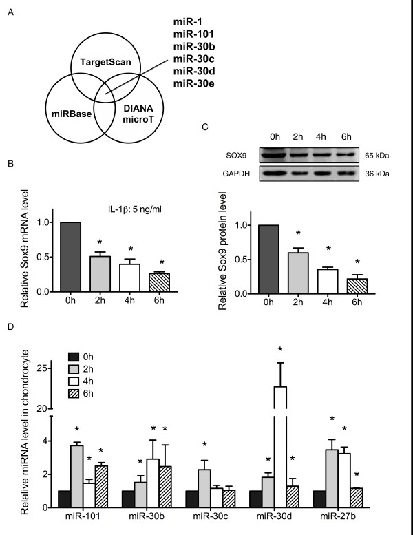
Introduction: Extracellular matrix (ECM) degradation leads to malfunction of the cartilage in osteoarthritis (OA). Inflammatory cytokine interleukin-1 beta (IL-1β) functions in ECM degradation and prevents ECM synthesis by down-regulating the key transcription factor, Sox9, and consequently inhibiting ECM gene expression. Evidence reveals that microRNAs (miRNA) have been associated with OA, but little is known of their function in chondrocyte ECM degradation. This study aimed to identify possible miRNAs that mediate IL-1β-induced down-regulation of Sox9 as well as its known down-stream genes, collagen type II and aggrecan.
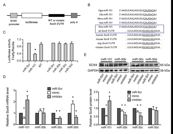
Methods: The miRNAs were predicted based on three classical databases. The expression levels of the predicted miRNAs were assessed in IL-1β stimulated chondrocytes by real-time PCR. A luciferase reporter was used to test the binding of the miRNAs to the 3' untranslated regions (3'UTR) of Sox9. The predicted miRNAs were transfected into chondrocytes to validate their relationship with Sox9. Functional analysis of the miRNAs on chondrocytes ECM degradation was performed at both the mRNA and protein levels after miRNA transfection and IL-1β treatment.
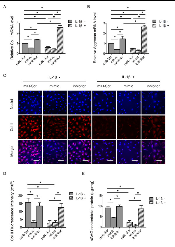
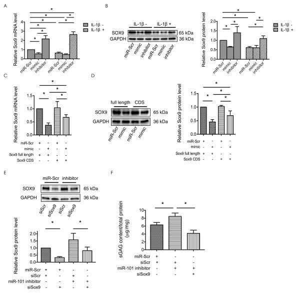
Results: Six miRNAs were predicted to target Sox9, and their expression in IL-1β-stimulated chondrocytes was revealed by real-time PCR. The luciferase reporter assay indicated that only miR-101 could bind to the 3'UTR of Sox9. The expression of Sox9 was likewise negatively regulated by miR-101 in rat chondrocytes. Functional analysis showed that miR-101 could aggravate chondrocyte ECM degradation, whereas miR-101 inhibition could reverse IL-1β-induced ECM degradation.
Conclusion: miR-101 participates in IL-1β-induced chondrocyte ECM degradation. Down-regulating miR-101 expression can prevent the IL-1β-induced ECM degradation in chondrocytes. miR-101 probably functions by directly targeting Sox9 mRNA.
Figures
Similar articles
-
Downregulation of miR-221-3p contributes to IL-1β-induced cartilage degradation by directly targeting the SDF1/CXCR4 signaling pathway.J Mol Med (Berl). 2017 Jun;95(6):615-627. doi: 10.1007/s00109-017-1516-6. Epub 2017 Feb 24. J Mol Med (Berl). 2017. PMID: 28236026
-
MicroRNA-30a promotes extracellular matrix degradation in articular cartilage via downregulation of Sox9.Cell Prolif. 2016 Apr;49(2):207-18. doi: 10.1111/cpr.12246. Epub 2016 Mar 10. Cell Prolif. 2016. PMID: 26969024 Free PMC article.
-
CircSERPINE2 weakens IL-1β-caused apoptosis and extracellular matrix degradation of chondrocytes by regulating miR-495/TGFBR2 axis.Biosci Rep. 2020 Nov 27;40(11):BSR20201601. doi: 10.1042/BSR20201601. Biosci Rep. 2020. PMID: 33094798 Free PMC article.
-
Illustrating the interplay between the extracellular matrix and microRNAs.Int J Exp Pathol. 2014 Jun;95(3):158-80. doi: 10.1111/iep.12079. Epub 2014 Apr 25. Int J Exp Pathol. 2014. PMID: 24761792 Free PMC article. Review.
-
miRNAs regulate expression and function of extracellular matrix molecules.Matrix Biol. 2013 Mar 11;32(2):74-85. doi: 10.1016/j.matbio.2012.11.003. Epub 2012 Nov 15. Matrix Biol. 2013. PMID: 23159731 Free PMC article. Review.
Cited by
-
Silencing of miR-101 Prevents Cartilage Degradation by Regulating Extracellular Matrix-related Genes in a Rat Model of Osteoarthritis.Mol Ther. 2015 Aug;23(8):1331-1340. doi: 10.1038/mt.2015.61. Epub 2015 Apr 29. Mol Ther. 2015. PMID: 25921548 Free PMC article.
-
SOX9 and the many facets of its regulation in the chondrocyte lineage.Connect Tissue Res. 2017 Jan;58(1):2-14. doi: 10.1080/03008207.2016.1183667. Epub 2016 Apr 29. Connect Tissue Res. 2017. PMID: 27128146 Free PMC article. Review.
-
The complex landscape of microRNAs in articular cartilage: biology, pathology, and therapeutic targets.JCI Insight. 2018 Sep 6;3(17):e121630. doi: 10.1172/jci.insight.121630. eCollection 2018 Sep 6. JCI Insight. 2018. PMID: 30185670 Free PMC article. Review.
-
MiR-146b accelerates osteoarthritis progression by targeting alpha-2-macroglobulin.Aging (Albany NY). 2019 Aug 17;11(16):6014-6028. doi: 10.18632/aging.102160. Epub 2019 Aug 17. Aging (Albany NY). 2019. PMID: 31422941 Free PMC article.
-
Garcinol Suppresses IL-1β-Induced Chondrocyte Inflammation and Osteoarthritis via Inhibition of the NF-κB Signaling Pathway.Inflammation. 2019 Oct;42(5):1754-1766. doi: 10.1007/s10753-019-01037-7. Inflammation. 2019. PMID: 31201586
References
Publication types
MeSH terms
Substances
LinkOut - more resources
Full Text Sources
Research Materials
