Review
. 2013 Feb;13(2):317-29.
doi: 10.2174/156652413804810781.

Cell death and survival through the endoplasmic reticulum-mitochondrial axis

Affiliations
Review

Cell death and survival through the endoplasmic reticulum-mitochondrial axis

R Bravo-Sagua et al. Curr Mol Med. 2013 Feb.

Abstract

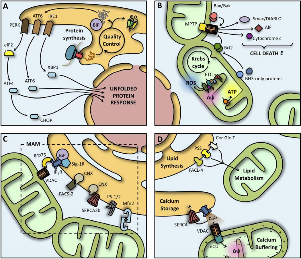
The endoplasmic reticulum has a central role in biosynthesis of a variety of proteins and lipids. Mitochondria generate ATP, synthesize and process numerous metabolites, and are key regulators of cell death. The architectures of endoplasmic reticulum and mitochondria change continually via the process of membrane fusion, fission, elongation, degradation, and renewal. These structural changes correlate with important changes in organellar function. Both organelles are capable of moving along the cytoskeleton, thus changing their cellular distribution. Numerous studies have demonstrated coordination and communication between mitochondria and endoplasmic reticulum. A focal point for these interactions is a zone of close contact between them known as the mitochondrial-associated endoplasmic reticulum membrane (MAM), which serves as a signaling juncture that facilitates calcium and lipid transfer between organelles. Here we review the emerging data on how communication between endoplasmic reticulum and mitochondria can modulate organelle function and determine cellular fate.


Conflict of interest statement

CONFLICT OF INTEREST

The authors confirm that this article content has no conflicts of interest.

Figures

Fig. 1
Fig. 1. ER and mitochondrial signaling cascades
(A) Accumulation of unfolded proteins in the ER lumen releases Bip and activates transmembrane sensors IRE1, PERK and ATF6, whose signaling recruits several transcription factors (XBP1, ATF4 and CHOP), leading to the activation of the Unfolded Protein Response genetic program. (B) Mitochondria perform essential metabolic reactions (Krebs cycle, ATP production, ROS generation), and are key regulators of cell death by receiving different inputs (e.g. Bcl-2 family proteins) and compartmentalizing potent inductors of apoptosis (AIF, cytochrome c and Smac/DIABLO). (C) Several proteins are compartmentalized in the Mitochondria-Associated Membranes, allowing a direct ER-mitochondria communication. Noteworthy is the participation of GRP75, forming a bridge between the Ca2+ channels IP3R and VDAC, while Calnexin (CNX) directly modulates the activity of SERCA2b. (D) The ER releases Ca2+ through specialized channels (IP3R), and pumps it back through SERCA. Mitochondria directly uptakes this Ca2+ through a series of channels (mCU and VDAC) driven by its transmembrane potential (Δψ). ER-mitochondria communication also allows the transfer of lipids between organelles at the MAM.
Fig. 2
Fig. 2. Life and death through the ER-mitochondria axis
(A) Severe ER stress induces pro-apoptotic signaling from the ER (IRE1 and PERK) that leads to mitochondrial Ca2+ overload. ER-directed mitochondrial fragmentation (via tBid and p20) preludes mitochondrial permeabilization and release of apoptotic factors (e.g. cytochrome C). (B) Non-lethal ER stress induces a microtubule-dependent spatial reorganization of ER and mitochondria that leads to enhanced organelle coupling and calcium transfer. Increased Ca2+ uptake produces a boost in mitochondrial bioenergetics and ATP production as an adaptative response. (C) The engagement of the UPR upon mitochondrial damage activates the mitophagy pathway, coordinating the elimination of dysfunctional organelles. (D) Disturbances in general homeostasis require a synchronic response from ER and mitochondria. While ER signaling protects mitochondrial integrity, mitochondria have important components for proper UPR signaling.

References

    1. Berridge MJ. The endoplasmic reticulum: a multifunctional signaling organelle. Cell Calcium. 2002;32:235–249. - PubMed
    1. Johnson AE, Haigh NG. The ER translocon and retrotranslocation: is the shift into reverse manual or automatic? Cell. 2000;102:709–712. - PubMed
    1. Ellgaard L, Helenius A. Quality control in the endoplasmic reticulum. Nat Rev Mol Cell Biol. 2003;4:181–191. - PubMed
    1. Ni M, Lee AS. ER chaperones in mammalian development and human diseases. FEBS Lett. 2007;581:3641–3651. - PMC - PubMed
    1. van Meer G, Voelker DR, Feigenson GW. Membrane lipids: where they are and how they behave. Nat Rev Mol Cell Biol. 2008;9:112–124. - PMC - PubMed

Publication types

LinkOut - more resources