

GLUT1 expression patterns in different Hodgkin lymphoma subtypes and progressively transformed germinal centers
- PMID: 23228169
- PMCID: PMC3537691
- DOI: 10.1186/1471-2407-12-586
GLUT1 expression patterns in different Hodgkin lymphoma subtypes and progressively transformed germinal centers
Abstract
Background: Increased glycolytic activity is a hallmark of cancer, allowing staging and restaging with 18F-fluorodeoxyglucose-positron-emission-tomography (PET). Since interim-PET is an important prognostic tool in Hodgkin lymphoma (HL), the aim of this study was to investigate the expression of proteins involved in the regulation of glucose metabolism in the different HL subtypes and their impact on clinical outcome.
Methods: Lymph node biopsies from 54 HL cases and reactive lymphoid tissue were stained for glucose transporter 1 (GLUT1), lactate dehydrogenase A (LDHA) and lactate exporter proteins MCT1 and MCT4. In a second series, samples from additional 153 HL cases with available clinical data were stained for GLUT1 and LDHA.
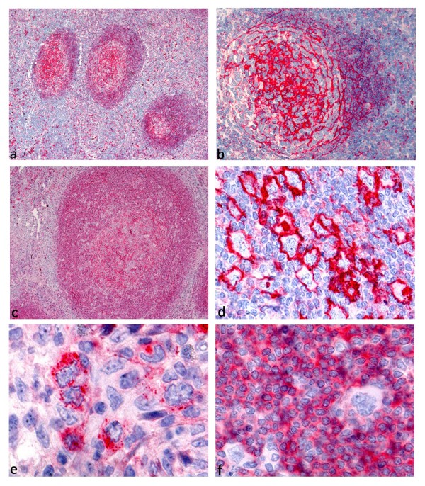
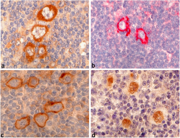
Results: Membrane bound GLUT1 expression was frequently observed in the tumor cells of HL (49% of all cases) but showed a broad variety between the different Hodgkin lymphoma subtypes: Nodular sclerosing HL subtype displayed a membrane bound GLUT1 expression in the Hodgkin-and Reed-Sternberg cells in 56% of the cases. However, membrane bound GLUT1 expression was more rarely observed in tumor cells of lymphocyte rich classical HL subtype (30%) or nodular lymphocyte predominant HL subtype (15%). Interestingly, in both of these lymphocyte rich HL subtypes as well as in progressively transformed germinal centers, reactive B cells displayed strong expression of GLUT1. LDHA, acting downstream of glycolysis, was also expressed in 44% of all cases. We evaluated the prognostic value of different GLUT1 and LDHA expression patterns; however, no significant differences in progression free or overall survival were found between patients exhibiting different GLUT1 or LDHA expression patterns. There was no correlation between GLUT1 expression in HRS cells and PET standard uptake values.
Conclusions: In a large number of cases, HRS cells in classical HL express high levels of GLUT1 and LDHA indicating glycolytic activity in the tumor cells. Although interim-PET is an important prognostic tool, a predictive value of GLUT1 or LDHA staining of the primary diagnostic biopsy could not be demonstrated. However, we observed GLUT1 expression in progressively transformed germinal centers and hyperplastic follicles, explaining false positive results in PET. Therefore, PET findings suggestive of HL relapse should always be confirmed by histology.
Figures
Similar articles
-
Hodgkin lymphoma: A complex metabolic ecosystem with glycolytic reprogramming of the tumor microenvironment.Semin Oncol. 2017 Jun;44(3):218-225. doi: 10.1053/j.seminoncol.2017.10.003. Epub 2017 Oct 10. Semin Oncol. 2017. PMID: 29248133 Free PMC article.
-
Glut1 and Glut3 expression in lymphoma and their association with tumor intensity on 18F-fluorodeoxyglucose positron emission tomography.Nucl Med Commun. 2009 Aug;30(8):594-601. doi: 10.1097/MNM.0b013e32832cc295. Nucl Med Commun. 2009. PMID: 19536037
-
Relationship between FDG uptake and expressions of glucose transporter type 1, type 3, and hexokinase-II in Reed-Sternberg cells of Hodgkin lymphoma.Oncol Res. 2009;17(7):331-7. doi: 10.3727/096504009787721177. Oncol Res. 2009. PMID: 19408578
-
The Hodgkin and Reed/Sternberg cell.Int J Biochem Cell Biol. 2005 Mar;37(3):511-7. doi: 10.1016/j.biocel.2003.10.025. Int J Biochem Cell Biol. 2005. PMID: 15618006 Review.
-
Hodgkin lymphoma: Pathology and biology.Semin Hematol. 2016 Jul;53(3):139-47. doi: 10.1053/j.seminhematol.2016.05.007. Epub 2016 May 13. Semin Hematol. 2016. PMID: 27496304 Review.
Cited by
-
Metabolic Function and Therapeutic Potential of CD147 for Hematological Malignancies: An Overview.Int J Mol Sci. 2024 Aug 23;25(17):9178. doi: 10.3390/ijms25179178. Int J Mol Sci. 2024. PMID: 39273126 Free PMC article. Review.
-
Dasatinib associated lymphadenopathy in a chronic myeloid leukemia patient: A case report.Medicine (Baltimore). 2020 Nov 6;99(45):e22791. doi: 10.1097/MD.0000000000022791. Medicine (Baltimore). 2020. PMID: 33157925 Free PMC article.
-
Interim PET-results for prognosis in adults with Hodgkin lymphoma: a systematic review and meta-analysis of prognostic factor studies.Cochrane Database Syst Rev. 2020 Jan 13;1(1):CD012643. doi: 10.1002/14651858.CD012643.pub3. Cochrane Database Syst Rev. 2020. PMID: 31930780 Free PMC article.
-
18F-Fluorodeoxyglucose positron emission tomography computed tomography detection threshold in follicular lymphoma: A case report.Medicine (Baltimore). 2017 Nov;96(47):e8705. doi: 10.1097/MD.0000000000008705. Medicine (Baltimore). 2017. PMID: 29381956 Free PMC article.
-
[Nodular lymphocyte-predominant Hodgkin's lymphoma and differential diagnoses].Pathologe. 2013 May;34(3):233-43. doi: 10.1007/s00292-013-1747-4. Pathologe. 2013. PMID: 23494280 Review. German.
References
-
- Küppers R, Rajewsky K, Zhao M, Simons G, Laumann R, Fischer R. et al.Hodgkin disease: Hodgkin and reed-Sternberg cells picked from histological sections show clonal immunoglobulin gene rearrangements and appear to be derived from B cells at various stages of development. Proc Natl Acad Sci USA. 1994;91:10962–10966. doi: 10.1073/pnas.91.23.10962. - DOI - PMC - PubMed
-
- Swerdlow SH, International Agency for Research on Cancer. World Health Organization. WHO classification of tumours of haematopoietic and lymphoid tissues. 4. Lyon, France: International Agency for Research on Cancer; 2008.
MeSH terms
Substances
LinkOut - more resources
Full Text Sources
Medical
Miscellaneous
