. 2013 Jun;84(2):335-44.
doi: 10.1016/j.ejpb.2012.11.023. Epub 2012 Dec 8.

Inhalation treatment of pulmonary fibrosis by liposomal prostaglandin E2

Affiliations

Inhalation treatment of pulmonary fibrosis by liposomal prostaglandin E2

Vera Ivanova et al. Eur J Pharm Biopharm. 2013 Jun.

Abstract

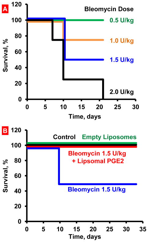
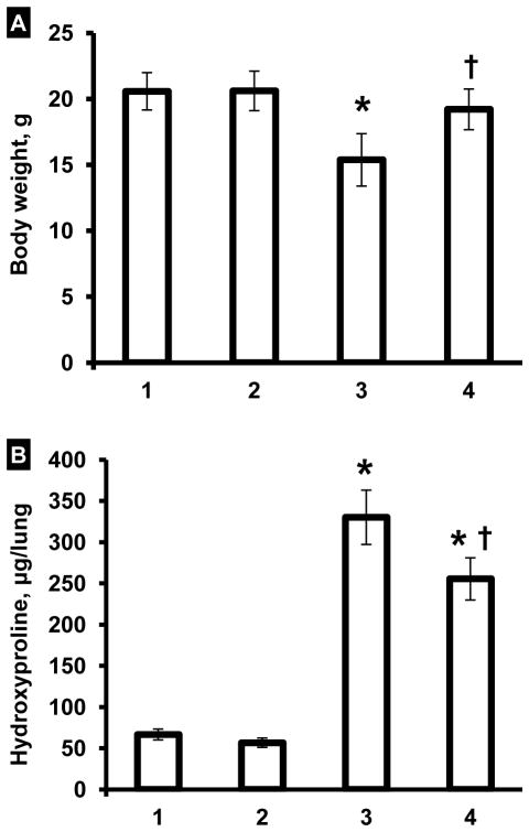
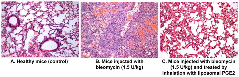
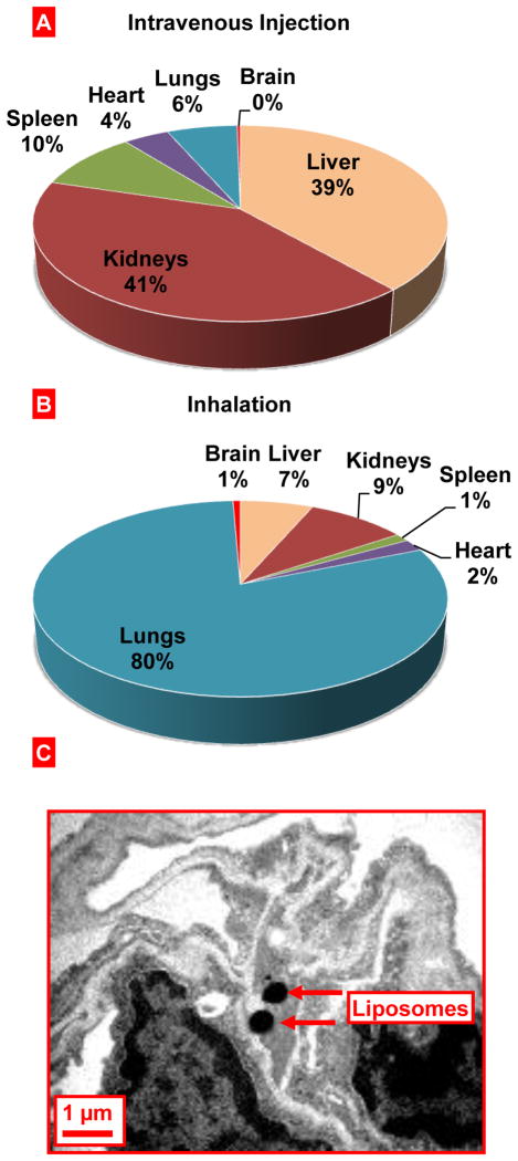
Idiopathic pulmonary fibrosis (IPF) is a chronic, progressive, and often fatal form of interstitial lung disease. We hypothesized that the local pulmonary delivery of prostaglandin E2 (PGE2) by liposomes can be used for the effective treatment of IPF. To test this hypothesis, we used a murine model of bleomycin-induced IPF to evaluate liposomal delivery of PGE2 topically to the lungs. Animal survival, body weight, hydroxyproline content in the lungs, lung histology, mRNA, and protein expression were studied. After inhalation delivery, liposomes accumulated predominately in the lungs. In contrast, intravenous administration led to the accumulation of liposomes mainly in kidney, liver, and spleen. Liposomal PGE2 prevented the disturbances in the expression of many genes associated with the development of IPF, substantially restricted inflammation and fibrotic injury in the lung tissues, prevented decrease in body weight, limited hydroxyproline accumulation in the lungs, and virtually eliminated mortality of animals after intratracheal instillation of bleomycin. In summary, our data provide evidence that pulmonary fibrosis can be effectively treated by the inhalation administration of liposomal form of PGE2 into the lungs. The results of the present investigations make the liposomal form of PGE2 an attractive drug for the effective inhalation treatment of idiopathic pulmonary fibrosis.


Figures

Figure 1
Figure 1
Survival of mice (Kaplan-Meier survival plot) after inhalation exposure to bleomycin. A – Selection of bleomycin dose. Mice were instilled intratracheally once with different concentrations of bleomycin. The dose of 1.5 U/kg that led to death of 50% of animals was selected. B – Inhalation treatment of mice with experimental lung fibrosis by liposomal PGE2 (Lip PGE2) prevents animal mortality. Inhalation of healthy mice with empty liposomes did not influence on animal survival. Lung fibrosis was induced by intratracheal instillation of 1.5 U/kg of bleomycin. Mice were treated with liposomal PGE2 twice a week for three weeks starting one day after the bleomycin administration.
Figure 2
Figure 2
Influence of inhalation treatment with liposomal PGE2 on body weight (A) and hydroxyproline content in the lungs (B). Lung fibrosis was induced by intratracheal instillation of 1.5 U/kg of bleomycin. Mice were treated with liposomal PGE2 twice a week for three weeks starting one day later after the bleomycin administration. At the end of treatment, lungs were harvested, homogenized and hydroxyproline content in the lungs was measured. 1 – Healthy mice (control); 2 – Healthy mice treated by inhalation with empty liposomes; 3 – Mice instilled with bleomycin (1.5 U/kg); 4 – Mice instilled with bleomycin (1.5 U/kg) and treated by inhalation with liposomal PGE2. Means ± S.D. are shown. *P < 0.05 when compared with control (1). P < 0.05 when compared with mice with fibrosis (2).
Figure 3
Figure 3
Lung histology. A- Healthy mice (control). B-Lung fibrosis was induced by intratracheal instillation of 1.5 U/kg of bleomycin. C-Mice were treated with inhalation of liposomal PGE2 within 3 weeks twice a week starting one day later after the bleomycin administration. At the end of experiment, lungs were harvested and fixed in 10% phosphate-buffered formalin. Samples were subsequently dehydrated and embedded in Paraplast®. Sections (5 μm) were cut, stained with hematoxylin and eosin. Representative images are shown (10X magnification).
Figure 4
Figure 4
Inhalation delivery of liposomes to mouse lungs. A, B - Relative tissue content of liposomes delivered to mice by intravenous instillation (A) or inhalation (B). Liposomal content was registered in organs 24 h after inhalation. C - Localization of liposomes in the mouse lung tissues after the inhalation delivery. Liposomes were labeled by osmium tetroxide and visualized by electron transmission microscopy.
Figure 5
Figure 5
Gene expression measured by the Quantitative Polymerase Chain Reaction (QPCR). The QPCR was performed using a standard Mouse Fibrosis RT Profiler PCR Array panel. Lung fibrosis was induced by intratracheal instillation of 1.5 U/kg of bleomycin. Mice were treated by inhalation delivery of liposomal PGE2 twice a week for three weeks starting one day later after the bleomycin administration. A – Mice instilled with bleomycin (1.5 U/kg); B – Mice instilled with bleomycin (1.5 U/kg) and treated by inhalation with liposomal PGE2.). Means ± SD are shown.
Figure 6
Figure 6
Expression of proteins (immunohistochemistry) in lung tissues. Representative images of tissue sections stained with antibodies against VEGF, CCL12, MMP3 and HIF1A proteins (10X magnification) and average expression of corresponding proteins. High intensity of the color indicates high protein concentration. Lung fibrosis was induced by intratracheal instillation of 1.5 U/kg of bleomycin. Mice were treated with liposomal PGE2 twice a week for three weeks starting one day later after the bleomycin administration. 1 - Healthy mice (control); 2-Mice instilled with bleomycin (1.5 U/kg); 3 - Mice instilled with bleomycin (1.5 U/kg) and treated by inhalation with liposomal PGE2. Means ± SD from are shown. *P < 0.05 when compared with healthy mice (control). P < 0.05 when compared with mice instilled with bleomycin.
Figure 7
Figure 7
Gene expression analyzed by RT-PCR. Representative images of gel electrophoresis of RT-PCR product and average expression of genes encoding hypoxia inducible factor 1α (HIF1A), von Hippel-Lindau (VHL) and β-actin (B-ACTIN, internal standard) proteins. 1 - Healthy mice (control); 2-Mice instilled with bleomycin (1.5 U/kg); 3 - Mice instilled with bleomycin (1.5 U/kg) and treated by inhalation with liposomal PGE2. Means ± SD from are shown. *P < 0.05 when compared with healthy mice (control). P < 0.05 when compared with mice instilled with bleomycin.

References

    1. American Thoracic Society/European Respiratory Society International Multidisciplinary Consensus Classification of the Idiopathic Interstitial Pneumonias. This joint statement of the American Thoracic Society (ATS), and the European Respiratory Society (ERS) was adopted by the ATS board of directors, June 2001 and by the ERS Executive Committee, June 2001. Am J Respir Crit Care Med. 2002;165:277–304. - PubMed
    1. Gharaee-Kermani M, Gyetko MR, Hu B, Phan SH. New insights into the pathogenesis and treatment of idiopathic pulmonary fibrosis: a potential role for stem cells in the lung parenchyma and implications for therapy. Pharm Res. 2007;24:819–841. - PubMed
    1. Sime PJ, O’Reilly KM. Fibrosis of the lung and other tissues: new concepts in pathogenesis and treatment. Clin Immunol. 2001;99:308–319. - PubMed
    1. Frankel SK, Schwarz MI. Update in idiopathic pulmonary fibrosis. Curr Opin Pulm Med. 2009;15:463–469. - PubMed
    1. Williams TJ, Wilson JW. Challenges in pulmonary fibrosis: 7--Novel therapies and lung transplantation. Thorax. 2008;63:277–284. - PubMed

Publication types

MeSH terms