

doi: 10.1016/j.jsb.2012.11.006.
Epub 2012 Dec 8.
Affinity grid-based cryo-EM of PKC binding to RACK1 on the ribosome
Affiliations
- PMID: 23228487
- PMCID: PMC3833090
- DOI: 10.1016/j.jsb.2012.11.006
Item in Clipboard
Affinity grid-based cryo-EM of PKC binding to RACK1 on the ribosome
J Struct Biol.
2013 Feb.
Abstract
Affinity grids (AG) are specialized EM grids that bind macromolecular complexes containing tagged proteins to obtain maximum occupancy for structural analysis through single-particle EM. In this study, utilizing AG, we show that His-tagged activated PKC βII binds to the small ribosomal subunit (40S). We reconstructed a cryo-EM map which shows that PKC βII interacts with RACK1, a seven-bladed β-propeller protein present on the 40S and binds in two different regions close to blades 3 and 4 of RACK1. This study is a first step in understanding the molecular framework of PKC βII/RACK1 interaction and its role in translation.
Copyright © 2012 Elsevier Inc. All rights reserved.
Figures
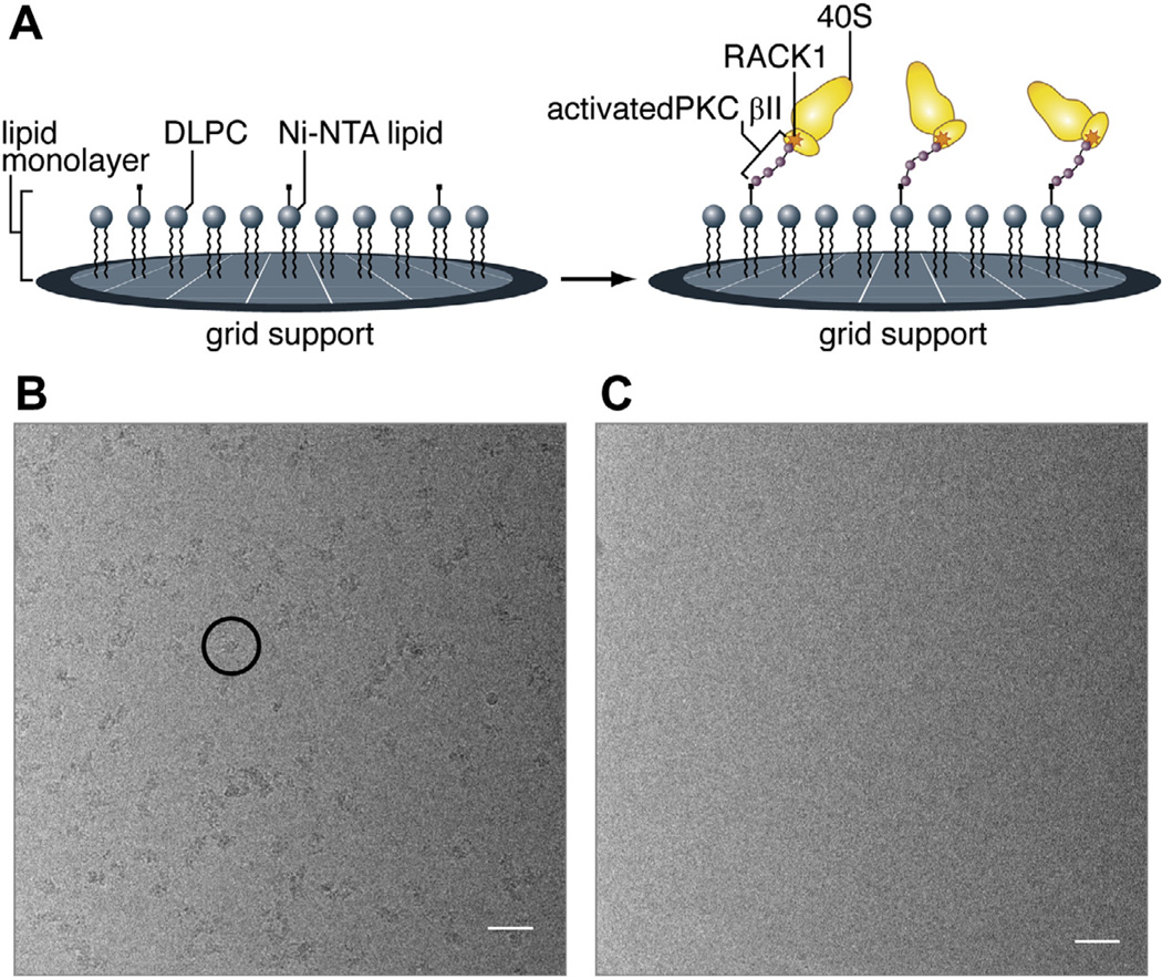
Affinity grid preparation of PKC-40S. (A) Schematic illustration of the affinity grid method and how PKC-40S associates on the grid surface. Micrographs of purified 40S subunits (rabbit reticulocytes) on Nickel-affinity grids (B) with and (C) without His-tagged PKC βII placed on affinity grids. The scale on the images is 500 Å. The black circle shows a representative of PKC-40S subunit.
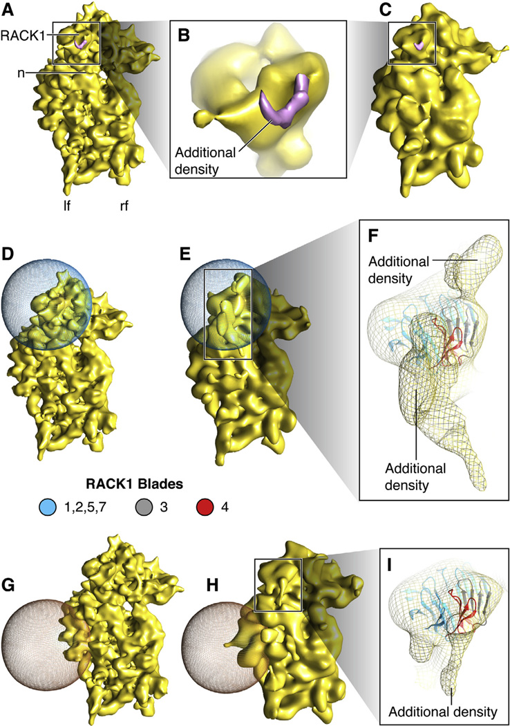
Density map of PKC-40S and Focused alignment. (A) Density map of PKC-40S at 11.4 Å. PKC-40S, is viewed from the solvent side. Small ribosomal subunit landmarks: n: neck; lf: left foot; rf: right foot, and RACK1. (B) Close-up showing rigid body docking of RACK1 in PKC-40S density map with additional density. (C) Density map from (A) displayed at same resolution as (E) and (H). (D and E) Focused alignment procedure performed to obtain partial densities of PKC βII near blades 3 and 4 of RACK1; the 3D mask is represented in blue mesh. (F) Close-up showing additional densities in contact with RACK1; the region highlighted in pink is suggested to interact with PKC βII (Grosso et al., 2008b; Ron et al., 1994). (G and H) Focused alignment procedure performed on another part of the 40S subunit, as control, showing very little additional density. (I) Close-up showing additional density generated close to RACK1. RACK1 model is taken from Homo sapiens (PDB ID: 3AOW).
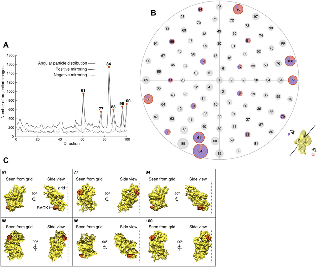
Orientation analysis in the PKC-40S data set. (A and B) Angular particle distribution plots for PKC-40S data set sorted in 100 view-directions. (A) The number of particles per direction, and in addition, positive and negative mirroring assigned during alignment are shown. (B) Each view on the starry sky plot is associated with two opposite orientation assignments, which we call P (in purple) and Q (in pink). For most views (circles in black) the same numbers of particles are facing in opposite directions, as indicated by the pie chart in a few randomly chosen examples. In contrast, the particles falling into the six overrepresented views (circled in orange) show a strong preference for one of the two directions. Cartoon at the bottom right explains the two opposite orientations, for an arbitrary view, giving rise to mirrored projections. (C) Relative proximity of RACK1 (in PKC-40S complex) to the cryo-grid in the six overrepresented orientations (as seen from the grid and the side-view, RACK1 is highlighted in orange).
References
-
- Ben-Shem A, Jenner L, Yusupova G, Yusupov M. Crystal structure of the eukaryotic ribosome. Science. 2010;330:1203–1209. - PubMed
-
- Frank J, Radermacher M, Penczek P, Zhu J, Li Y, et al. SPIDER and WEB: processing and visualization of images in 3D electron microscopy and related fields. J. Struct. Biol. 1996;116:190–199. - PubMed
-
- Grosso S, Volta V, Vietri M, Gorrini C, Marchisio PC, et al. Eukaryotic ribosomes host PKC activity. Biochem. Biophys. Res. Commun. 2008a;376:65–69. - PubMed
-
- Grosso S, Volta V, Sala LA, Vietri M, Marchisio PC, et al. PKCbetaII modulates translation independently from mTOR and through RACK1. Biochem. J. 2008b;415:77–85. - PubMed
Publication types
MeSH terms
Substances
Grants and funding
LinkOut - more resources
Full Text Sources
Other Literature Sources
