

β-amyloid dynamics in human plasma
- PMID: 23229043
- PMCID: PMC3808092
- DOI: 10.1001/archneurol.2012.18107
β-amyloid dynamics in human plasma
Abstract
Objectives: To investigate dynamic changes in human plasma β-amyloid (Aβ) concentrations, evaluate the effects of aging and amyloidosis on these dynamics, and determine their correlation with cerebrospinal fluid (CSF) Aβ concentrations.
Design: A repeated plasma and CSF sampling study.
Setting: The Washington University School of Medicine in St Louis, Missouri.
Participants: Older adults with amyloid deposition (Amyloid+), age-matched controls without amyloid deposition (Amyloid-), and younger normal controls (YNCs) were enrolled for the study.
Main outcome measures: Hourly measurements of plasma Aβ were compared between groups by age and amyloidosis. Plasma Aβ and CSF Aβ concentrations were compared for correlation, linear increase, and circadian patterns.
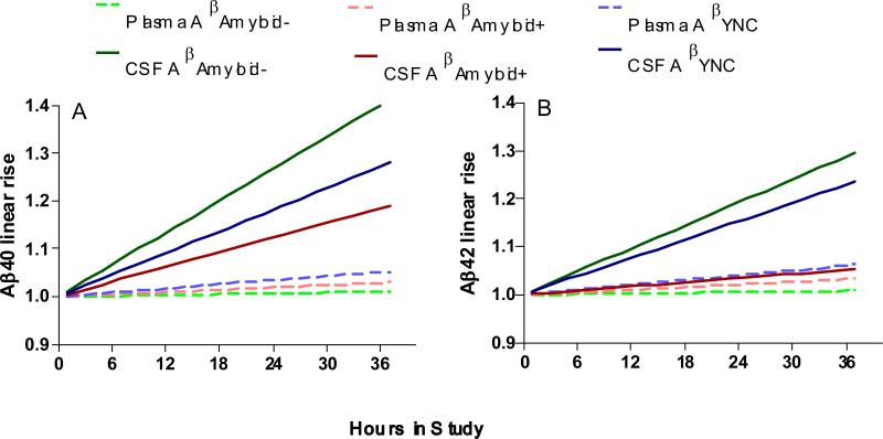
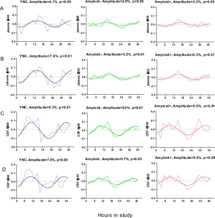
Results: Circadian patterns were observed in plasma Aβ, with diminished amplitudes with aging. Linear increase of Aβ was only observed for CSF Aβ in the YNC and Amyloid- groups, but not in the Amyloid+ group. No linear increase was observed for plasma Aβ. No significant correlations were found between plasma and CSF Aβ concentrations.
Conclusions: Plasma Aβ, like CSF, demonstrates a circadian pattern that is reduced in amplitude with increasing age but is unaffected by amyloid deposition. However, we found no evidence that plasma and CSF Aβ concentrations were related on an hourly or individual basis.
Figures
Similar articles
-
Effects of age and amyloid deposition on Aβ dynamics in the human central nervous system.Arch Neurol. 2012 Jan;69(1):51-8. doi: 10.1001/archneurol.2011.235. Epub 2011 Sep 12. Arch Neurol. 2012. PMID: 21911660 Free PMC article.
-
Associations Between β-Amyloid Kinetics and the β-Amyloid Diurnal Pattern in the Central Nervous System.JAMA Neurol. 2017 Feb 1;74(2):207-215. doi: 10.1001/jamaneurol.2016.4202. JAMA Neurol. 2017. PMID: 27992627 Free PMC article.
-
Age-dependent inverse correlations in CSF and plasma amyloid-β(1-42) concentrations prior to amyloid plaque deposition in the brain of 3xTg-AD mice.Sci Rep. 2016 Feb 2;6:20185. doi: 10.1038/srep20185. Sci Rep. 2016. PMID: 26830653 Free PMC article.
-
Is plasma amyloid-beta a reliable biomarker for Alzheimer's disease?Recent Pat CNS Drug Discov. 2008 Jun;3(2):109-11. doi: 10.2174/157488908784534595. Recent Pat CNS Drug Discov. 2008. PMID: 18537770 Review.
-
Cerebrospinal fluid Abeta40 and Abeta42: natural course and clinical usefulness.Front Biosci. 2002 Apr 1;7:d997-1006. doi: 10.2741/A826. Front Biosci. 2002. PMID: 11897565 Review.
Cited by
-
Candidate inflammatory biomarkers display unique relationships with alpha-synuclein and correlate with measures of disease severity in subjects with Parkinson's disease.J Neuroinflammation. 2017 Aug 18;14(1):164. doi: 10.1186/s12974-017-0935-1. J Neuroinflammation. 2017. PMID: 28821274 Free PMC article.
-
A randomized, controlled clinical trial of plasma exchange with albumin replacement for Alzheimer's disease: Primary results of the AMBAR Study.Alzheimers Dement. 2020 Oct;16(10):1412-1425. doi: 10.1002/alz.12137. Epub 2020 Jul 27. Alzheimers Dement. 2020. PMID: 32715623 Free PMC article. Clinical Trial.
-
Circadian regulation of membrane physiology in neural oscillators throughout the brain.Eur J Neurosci. 2020 Jan;51(1):109-138. doi: 10.1111/ejn.14343. Epub 2019 Jan 29. Eur J Neurosci. 2020. PMID: 30633846 Free PMC article. Review.
-
Plasma Amyloid Concentration in Alzheimer's Disease: Performance of a High-Throughput Amyloid Assay in Distinguishing Alzheimer's Disease Cases from Controls.J Alzheimers Dis. 2020;74(4):1285-1294. doi: 10.3233/JAD-200046. J Alzheimers Dis. 2020. PMID: 32176645 Free PMC article.
-
Alzheimer's Disease Diagnosis Using Misfolding Proteins in Blood.Dement Neurocogn Disord. 2020 Mar;19(1):1-18. doi: 10.12779/dnd.2020.19.1.1. Epub 2020 Mar 6. Dement Neurocogn Disord. 2020. PMID: 32174051 Free PMC article. Review.
References
-
- Aronson MK, Ooi WL, Geva DL, Masur D, Blau A, Frishman W. Dementia. Age-dependent incidence, prevalence, and mortality in the old old. Arch Intern Med. 1991 May;151(5):989–992. - PubMed
-
- Hardy J, Selkoe DJ. The amyloid hypothesis of Alzheimer's disease: progress and problems on the road to therapeutics. Science. 2002 Jul 19;297(5580):353–356. - PubMed
-
- Motter R, Vigo-Pelfrey C, Kholodenko D, et al. Reduction of beta-amyloid peptide42 in the cerebrospinal fluid of patients with Alzheimer's disease. Ann Neurol. 1995 Oct;38(4):643–648. - PubMed
-
- Galasko D, Chang L, Motter R, et al. High cerebrospinal fluid tau and low amyloid beta42 levels in the clinical diagnosis of Alzheimer disease and relation to apolipoprotein E genotype. Arch Neurol. 1998 Jul;55(7):937–945. - PubMed
Publication types
MeSH terms
Substances
Grants and funding
- P50 AG05681-22/AG/NIA NIH HHS/United States
- R-01-NS065667/NS/NINDS NIH HHS/United States
- R01 NS065667/NS/NINDS NIH HHS/United States
- P01 AG026276/AG/NIA NIH HHS/United States
- UL1 RR024992/RR/NCRR NIH HHS/United States
- UL1 TR000448/TR/NCATS NIH HHS/United States
- P01 AG03991-22/AG/NIA NIH HHS/United States
- K23 AG 03094601/AG/NIA NIH HHS/United States
- P01 AG003991/AG/NIA NIH HHS/United States
- P50 AG005681/AG/NIA NIH HHS/United States
- K08 AG027091-01/AG/NIA NIH HHS/United States
- K23 AG030946/AG/NIA NIH HHS/United States
- KL2 RR024994/RR/NCRR NIH HHS/United States
- K08 AG027091/AG/NIA NIH HHS/United States
LinkOut - more resources
Full Text Sources
Other Literature Sources
Medical
