

Three-dimensional images contribute to the diagnosis of mucous retention cyst in maxillary sinus
- PMID: 23229251
- PMCID: PMC3548636
- DOI: 10.4317/medoral.18141
Three-dimensional images contribute to the diagnosis of mucous retention cyst in maxillary sinus
Abstract
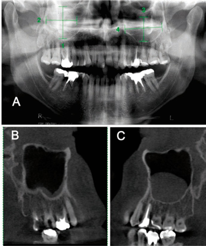
Objective: To evaluate the detection of mucous retention cyst of maxillary sinus (MRCMS) using panoramic radiography and cone beam computed tomography (CBCT).
Study design: A digital database with 6,000 panoramic radiographs was reviewed for MRCMS. Suggestive images of MRCMS were detected on 185 radiographs, and patients were located and invited to return for follow-up. Thirty patients returned, and control panoramic radiographs were obtained 6 to 46 months after the initial radiograph. When MRCMS was found on control radiographs, CBCT scans were obtained. Cysts were measured and compared on radiographs and scans. The Wilcoxon, Spearman and Kolmorogov-Smirnov tests were used for statistical analysis. The level of significance was set at 5%.
Results: There were statistically significant differences between the two methods (p<0.05): 23 MRCMS detected on panoramic radiographs were confirmed by CBCT, but 5 MRCMS detected on CBCT images had not been identified by panoramic radiography. Eight MRCMS detected on control radiographs were not confirmed by CBCT. MRCMS size differences from initial to control panoramic radiographs and CBCT scans were not statistically significant (p= 0.617 and p= 0.626). The correlation between time and MRCMS size differences was not significant (r = -0.16, p = 0.381).
Conclusion: CBCT scanning detect MRCMS more accurately than panoramic radiography.
Figures


Similar articles
-
Do CBCT scans alter surgical treatment plans? Comparison of preoperative surgical diagnosis using panoramic versus cone-beam CT images.J Craniomaxillofac Surg. 2016 Oct;44(10):1700-1705. doi: 10.1016/j.jcms.2016.07.025. Epub 2016 Aug 3. J Craniomaxillofac Surg. 2016. PMID: 27567358
-
Comparative assessment of panoramic radiography and CBCT imaging for radiodiagnostics in the posterior maxilla.Clin Oral Investig. 2014 Jan;18(1):293-300. doi: 10.1007/s00784-013-0963-x. Epub 2013 Mar 24. Clin Oral Investig. 2014. PMID: 23525890
-
The significance of cone beam computed tomography for the visualization of anatomical variations and lesions in the maxillary sinus for patients hoping to have dental implant-supported maxillary restorations in a private dental office in Japan.Head Face Med. 2014 May 28;10:20. doi: 10.1186/1746-160X-10-20. Head Face Med. 2014. PMID: 24884983 Free PMC article.
-
Performance of panoramic radiography compared with computed tomography in the evaluation of pathological changes in the maxillary sinuses: a systematic review and meta-analysis.Dentomaxillofac Radiol. 2023 Jul;52(5):20230067. doi: 10.1259/dmfr.20230067. Epub 2023 May 16. Dentomaxillofac Radiol. 2023. PMID: 37192021 Free PMC article.
-
Comparison of incidental findings on cone beam computed tomographic and 2-dimensional images.Gen Dent. 2023 Jul-Aug;71(4):64-71. Gen Dent. 2023. PMID: 37358586 Review.
Cited by
-
Frequency of Maxillary Sinus Mucous Retention Cysts in a Central Brazilian Population.J Dent (Shiraz). 2015 Sep;16(3):169-74. J Dent (Shiraz). 2015. PMID: 26331145 Free PMC article.
-
What is the frequency of anatomical variations and pathological findings in maxillary sinuses among patients subjected to maxillofacial cone beam computed tomography? A systematic review.Med Oral Patol Oral Cir Bucal. 2017 Jul 1;22(4):e400-e409. doi: 10.4317/medoral.21456. Med Oral Patol Oral Cir Bucal. 2017. PMID: 28578369 Free PMC article.
-
Differential protein expression in the secretory fluids of maxillary sinusitis and maxillary retention cyst.Eur Arch Otorhinolaryngol. 2017 Jan;274(1):215-222. doi: 10.1007/s00405-016-4167-2. Epub 2016 Jul 15. Eur Arch Otorhinolaryngol. 2017. PMID: 27422628
-
[Imaging classification diagnosis and maxillary sinus floor augmentation of maxillary sinus cystic lesions].Hua Xi Kou Qiang Yi Xue Za Zhi. 2019 Oct 1;37(5):457-462. doi: 10.7518/hxkq.2019.05.001. Hua Xi Kou Qiang Yi Xue Za Zhi. 2019. PMID: 31721489 Free PMC article. Chinese.
-
Evaluation of Mucous Retention Cyst Prevalence on Digital Panoramic Radiographs in the Local Population of Iran.Radiol Res Pract. 2022 Aug 8;2022:8650027. doi: 10.1155/2022/8650027. eCollection 2022. Radiol Res Pract. 2022. PMID: 35978990 Free PMC article.
References
-
- Myall RW, Eastep PB, Silver JG. Mucous retention cysts of the maxillary antrum. J Am Dent Assoc. 1974;89:1338–42. - PubMed
-
- Halstead CL. Mucosal cysts of the maxillary sinus: report of 75 cases. J Am Dent Assoc. 1973;87:1435–41. - PubMed
-
- Allard RH, van der Kwast WA, van der Waal I. Mucosal antral cysts. Review of the literature and report of a radiographic survey. Oral Surg Oral Med Oral Pathol. 1981;51:2–9. - PubMed
-
- Casamassimo PS, Lilly GE. Mucosal cysts of the maxillary sinus: a clinical and radiographic study. Oral Surg Oral Med Oral Pathol. 1980;50:282–6. - PubMed
-
- Gothberg KA, Little JW, King DR, Bean LR. A clinical study of cysts arising from mucosa of the maxillary sinus. Oral Surg Oral Med Oral Pathol. 1976;41:52–8. - PubMed
Publication types
MeSH terms
LinkOut - more resources
Full Text Sources
Other Literature Sources
