

Comparison of the value of PCNA and Ki-67 as markers of cell proliferation in ameloblastic tumors
- PMID: 23229269
- PMCID: PMC3613329
- DOI: 10.4317/medoral.18573
Comparison of the value of PCNA and Ki-67 as markers of cell proliferation in ameloblastic tumors
Abstract
Objectives: The aim of this study was to compare among PCNAand Ki-67 as the most reliable immunohistochemical marker for evaluating cell proliferation in ameloblastic tumors.
Study design: Observational, retrospective, and descriptive study of a large series of ameloblastic tumors, composed of 161 ameloblastomas and four ameloblastic carcinomas, to determine and compare PCNA and Ki-67 expression using immunohistochemistry techniques.
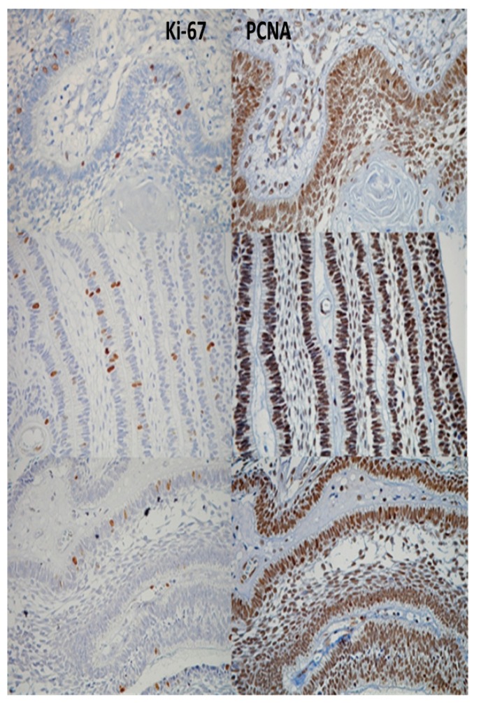
Results: When analyzing Ki-67 positivity, the desmoplastic ameloblastoma demonstrated a significantly lower proliferation rate (1.9%) compared with the solid/multicystic and unicystic ameloblastomas and ameloblastic carcinomas (p<0.05), whereas the ameloblastic carcinomas displayed a significantly higher rate compared with all of the other ameloblastomas (48.7%) (p<0.05). When analyzing cell proliferation with PCNA, we found significant differences only between the ameloblastic carcinomas (93.3%) and the desmoplastic ameloblastomas (p<0.05). When differences between the immunopositivity for PCNA and Ki-67 were compared, the percentages were higher for PCNA in all types of ameloblastomas and ameloblastic carcinomas. In all cases, the percentages were greater than 80%, whereas the immunopositivity for Ki-67 was significantly lower; for example, the ameloblastic carcinoma expressed the highest positivity and only reached 48.7%, compared to 93.3% when we used PCNA.
Conclusions: In the present study, when we used the proliferation cell marker Ki-67, the percentages of positivity were more specific and varied among the different types of ameloblastomas, suggesting that Ki-67 is a more specific marker for the proliferation of ameloblastic tumor cells.
Figures
References
-
- Bishop JM. The molecular genetics of cancer. Science. 1987;235:305–11. - PubMed
-
- Tumuluri V, Thomas GA, Fraser IS. Analysis of the Ki-67 antigen at the invasive tumour front of human oral squamous cell carcinoma. J Oral Pathol Med. 2002;31:598–604. - PubMed
-
- Scully C, Field JK, Tanzawa H. Genetic aberrations in oral or head and neck squamous cell carcinoma (SCCHN): 1. Carcinogen metabolism, DNA repair and cell cycle control. Oral Oncol. 2000 ;36:256–63. - PubMed
-
- Gerdes J, Schwab U, Lemke H, Stein H. Production of a mouse monoclonal antibody reactive with a human nuclear antigen associated with cell proliferation. Int J Cancer. 1983;31:13–20. - PubMed
Publication types
MeSH terms
Substances
LinkOut - more resources
Full Text Sources
Other Literature Sources
Medical
Miscellaneous
