. 2013 Feb;23(2):213-24.
doi: 10.1038/cr.2012.170. Epub 2012 Dec 11.

Intermolecular recognition revealed by the complex structure of human CLOCK-BMAL1 basic helix-loop-helix domains with E-box DNA

Affiliations

Intermolecular recognition revealed by the complex structure of human CLOCK-BMAL1 basic helix-loop-helix domains with E-box DNA

Zixi Wang et al. Cell Res. 2013 Feb.

Abstract

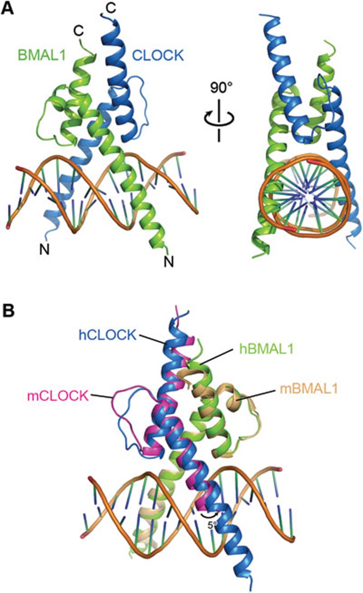
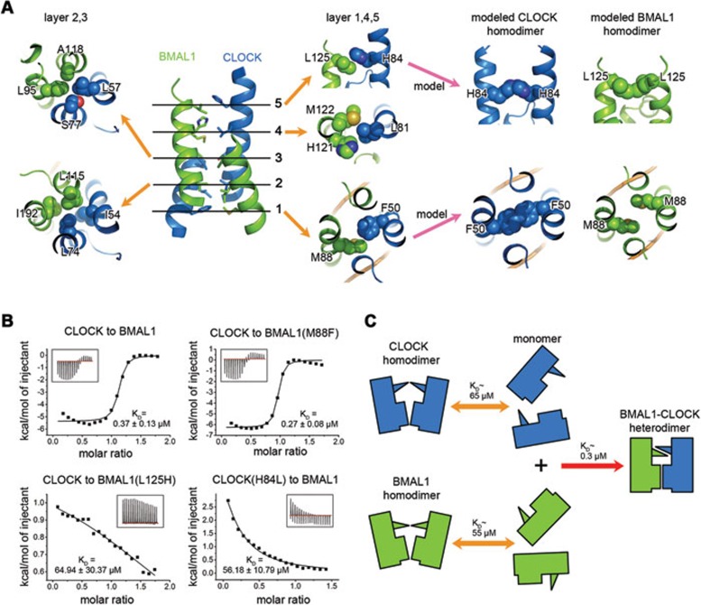
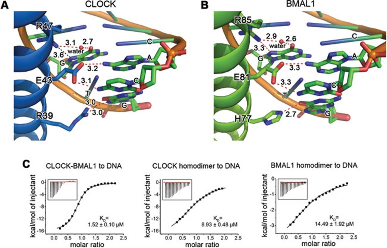
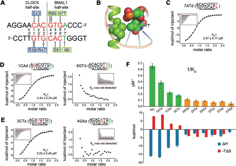
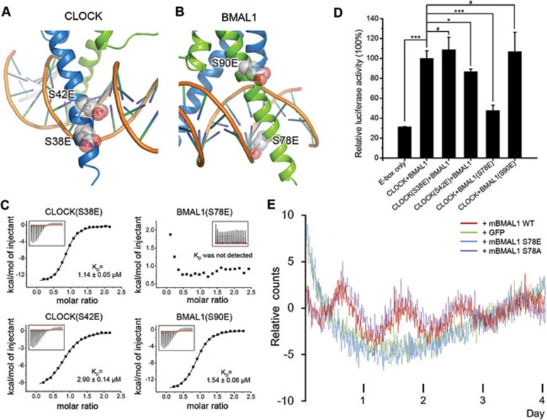
CLOCK (circadian locomotor output cycles kaput) and BMAL1 (brain and muscle ARNT-like 1) are both transcription factors of the circadian core loop in mammals. Recently published mouse CLOCK-BMAL1 bHLH (basic helix-loop-helix)-PAS (period-ARNT-single-minded) complex structure sheds light on the mechanism for heterodimer formation, but the structural details of the protein-DNA recognition mechanisms remain elusive. Here we have elucidated the crystal structure of human CLOCK-BMAL1 bHLH domains bound to a canonical E-box DNA. We demonstrate that CLOCK and BMAL1 bHLH domains can be mutually selected, and that hydrogen-bonding networks mediate their E-box recognition. We identified a hydrophobic contact between BMAL1 Ile80 and a flanking thymine nucleotide, suggesting that CLOCK-BMAL1 actually reads 7-bp DNA and not the previously believed 6-bp DNA. To find potential non-canonical E-boxes that could be recognized by CLOCK-BMAL1, we constructed systematic single-nucleotide mutations on the E-box and measured their relevant affinities. We defined two non-canonical E-box patterns with high affinities, AACGTGA and CATGTGA, in which the flanking A7-T7' base pair is indispensable for recognition. These results will help us to identify functional CLOCK-BMAL1-binding sites in vivo and to search for clock-controlled genes. Furthermore, we assessed the inhibitory role of potential phosphorylation sites in bHLH regions. We found that the phospho-mimicking mutation on BMAL1 Ser78 could efficiently block DNA binding as well as abolish normal circadian oscillation in cells. We propose that BMAL1 Ser78 should be a key residue mediating input signal-regulated transcriptional inhibition for external cues to entrain the circadian clock by kinase cascade.


Figures

Figure 1
Figure 1
Overall structure. (A) Heterodimeric structure of human CLOCK-BMAL1 bHLH domains with DNA. CLOCK is colored blue and BMAL1 is colored green. (B) Structural alignment of human CLOCK-BMAL1 bHLH domains and the counterpart of mouse (PDB: 4F3L).
Figure 2
Figure 2
The mutual recognition mechanism between CLOCK and BMAL1 bHLH domains. (A) The four-helix bundle composing the heterodimer interface can be divided into five layers. In layers 2 and 3, the residues in symmetric positions are the same (except CLOCK S77 and BMAL1 A118), so they do not confer the selectivity for the heterodimer. In the modeled homodimeric structures, CLOCK Phe50 in layer 1 and CLOCK His84, BMAL1 Leu125 in layer 5 have steric clash. (B) ITC measurements for the titration of the CLOCK bHLH domain into the BMAL1 bHLH domain. Mutated residues are indicated in subtitles. KD ± s.d. is shown. Homodimer-mimicking mutations in layer 5 (BMAL1 L125H and CLOCK H84L) greatly diminish the interaction. Homodimer-mimicking mutation in layer 1 (BMAL1 M88F) has no observable effect on mutual selection. (C) A proposed mechanistic model for mutual recognition. In the heterodimeric structure, the two recognition residues (CLOCK His84 and BMAL1 Leu125) are well stacked, thus leading a shift of the equilibrium to the heterodimer formation.
Figure 3
Figure 3
The recognition mechanism of CLOCK-BMAL1 bHLH domains to canonical E-box. (A) Detailed interactions between CLOCK and E-box. (B) Detailed interactions between BMAL1 and E-box. The red dashed lines represent the hydrogen-bonding contacts and the black dashed line represents the distance between two atoms. The bond lengths or distance are shown in Angstroms (Å). (C) ITC measurements of the binding affinities between hetero-/homodimeric proteins and the E-box DNA. bHLH domain proteins were titrated into a 16-bp blunt-ended double-stranded DNA that contains a canonical E-box in the center (Sequence: AGGAACACGTGACCCA. It is designated as WT DNA. E-box is underlined). Although the CLOCK bHLH homodimer has a higher affinity than BMAL1, both homodimers have a much lower affinity than heterodimer. KD ± s.d. is shown.
Figure 4
Figure 4
CLOCK and BMAL1 read DNA bases asymmetrically. (A) A schematic recognition diagram of CLOCK-BMAL1 to DNA in the crystal structure. Residues that participate in DNA recognition are labeled by a blue rectangle in CLOCK and a green rectangle in BMAL1. E-box is in red. Flanking base pairs that are recognized by BMAL1 is colored green. Black arrows represent hydrogen bonds. Lower-case 'w' means water molecule. Zebra triangle represents hydrophobic contact. DNA is divided into the CLOCK half-site and the BMAL1 half-site by a dashed line. (B) A close view of the hydrophobic contact between BMAL1 Ile80 and flanking T7′. (C) Titration of CLOCK-BMAL1 bHLH heterodimer into a 16 bp canonical E-box DNA with flanking T7′ mutated (7ATd, ACACGTGT). (D) CLOCK-BMAL1 binds to 1CAd (AAACGTGA) but not 6GTd (ACACGTTA). (E) CLOCK-BMAL1 binds to 3CTz (ACATGTGA) but not 4GAz (ACACATGA). The core DNA sequences used in titration are shown in subtitles and the E-box is underlined. Mutated base pairs are colored red, and the KD ± s.d. is shown. (F) The A2 or T5 site single-mutated E-box DNA molecules have much lower binding affinities and different thermodynamic profiles. 1/KD ± s.d. is shown. The core sequences of the mutated DNA are 2AGz (ACGCGTGA), 2ATd (ACTCGTGA), 2ACd (ACCCGTGA), 5TCz (ACACGCGA), 5TAd (ACACGAGA), 5TGd (ACACGGGA). See Supplementary information, Table S1 for full-length DNA used in ITC measurements.
Figure 5
Figure 5
Potential phosphorylation sites in basic regions and their effects on DNA binding. (A) Modeled phospho-mimicking mutations in CLOCK basic region. (B) Modeled phospho-mimicking mutations in BMAL1 basic region. (C) Effects of phospho-mimicking mutations on DNA binding. CLOCK-BMAL1 bHLH domains carrying the indicated mutations were titrated into WT E-box DNA. KD ± s.d. is shown. Only the BMAL1 S78E mutation effectively blocks DNA binding. (D) Luciferase reporter assay demonstrates that BMAL1 S78E can inhibit DNA binding in vivo, thus diminishing the transcriptional activity of CLOCK-BMAL1. Data were represented as mean ± s.d. of three repeats in one assay. P value was calculated by Student's t-test. ***P< 0.001; *P< 0.05; #P> 0.1. (E) Real-time whole-cell luciferase assay. mBMAL1 WT, mBMAL1 S78A, mBMAL1 S78E and GFP genes were transfected into mouse mBMAL1−/− mPer2Luciferase fibroblast cells, respectively. Real-time expression of the fusion protein mPER2-luciferase was monitored for 4 days. Red, purple, blue and green lines represent the luminescent signal using mBMAL1 WT, mBMAL1 S78A, mBMAL1 S78E and GFP as rescue genes, respectively.

References

    1. Panda S, Hogenesch JB, Kay SA. Circadian rhythms from flies to human. Nature. 2002;417:329–335. - PubMed
    1. Reppert SM, Weaver DR. Coordination of circadian timing in mammals. Nature. 2002;418:935–941. - PubMed
    1. Green CB, Takahashi JS, Bass J. The meter of metabolism. Cell. 2008;134:728–742. - PMC - PubMed
    1. Maury E, Ramsey KM, Bass J. Circadian rhythms and metabolic syndrome: from experimental genetics to human disease. Circ Res. 2010;106:447–462. - PMC - PubMed
    1. Dunlap JC. Molecular bases for circadian clocks. Cell. 1999;96:271–290. - PubMed

Publication types