. 2013 Feb 15;288(7):4799-809.
doi: 10.1074/jbc.M112.432781. Epub 2012 Dec 10.

Understanding how noncatalytic carbohydrate binding modules can display specificity for xyloglucan

Affiliations

Understanding how noncatalytic carbohydrate binding modules can display specificity for xyloglucan

Ana S Luís et al. J Biol Chem. .

Abstract

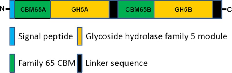
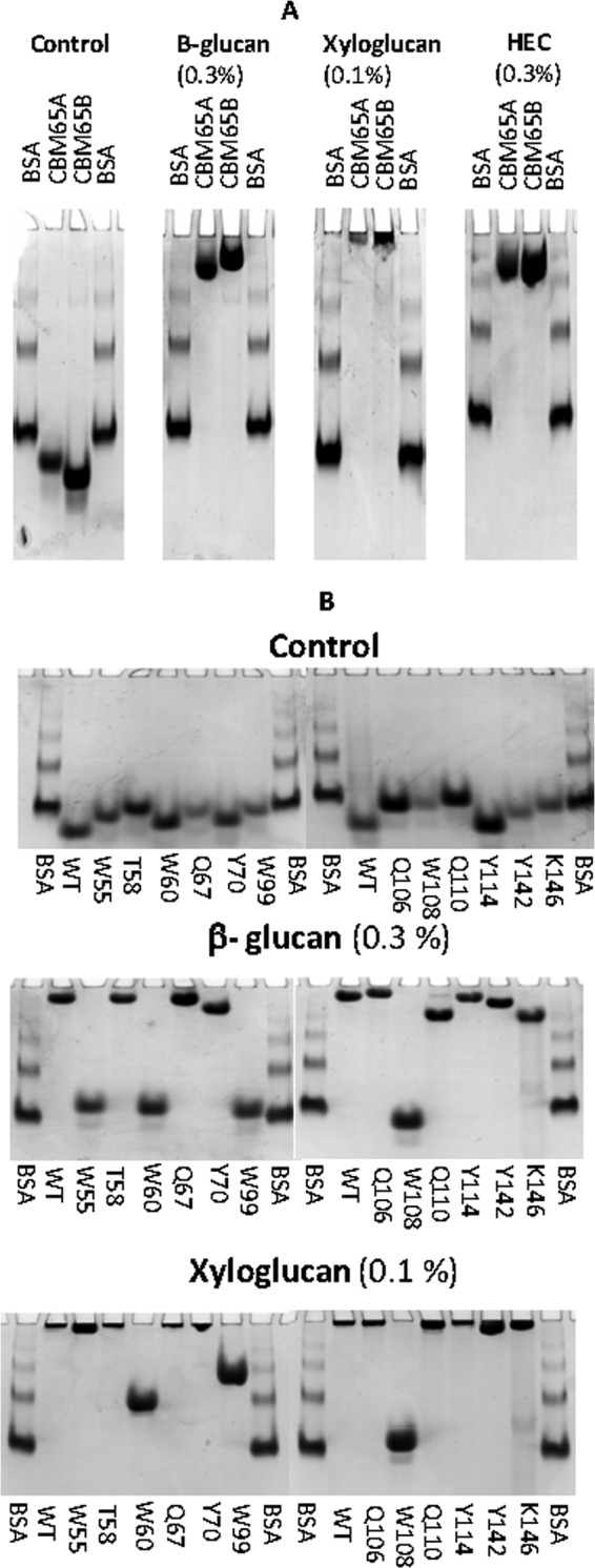
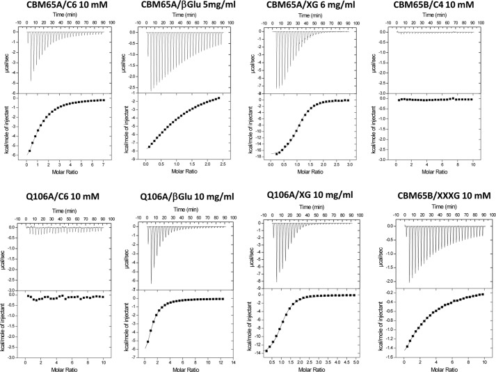
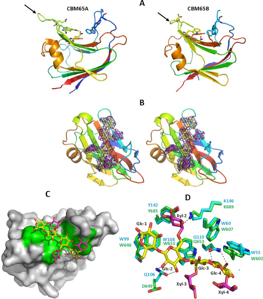
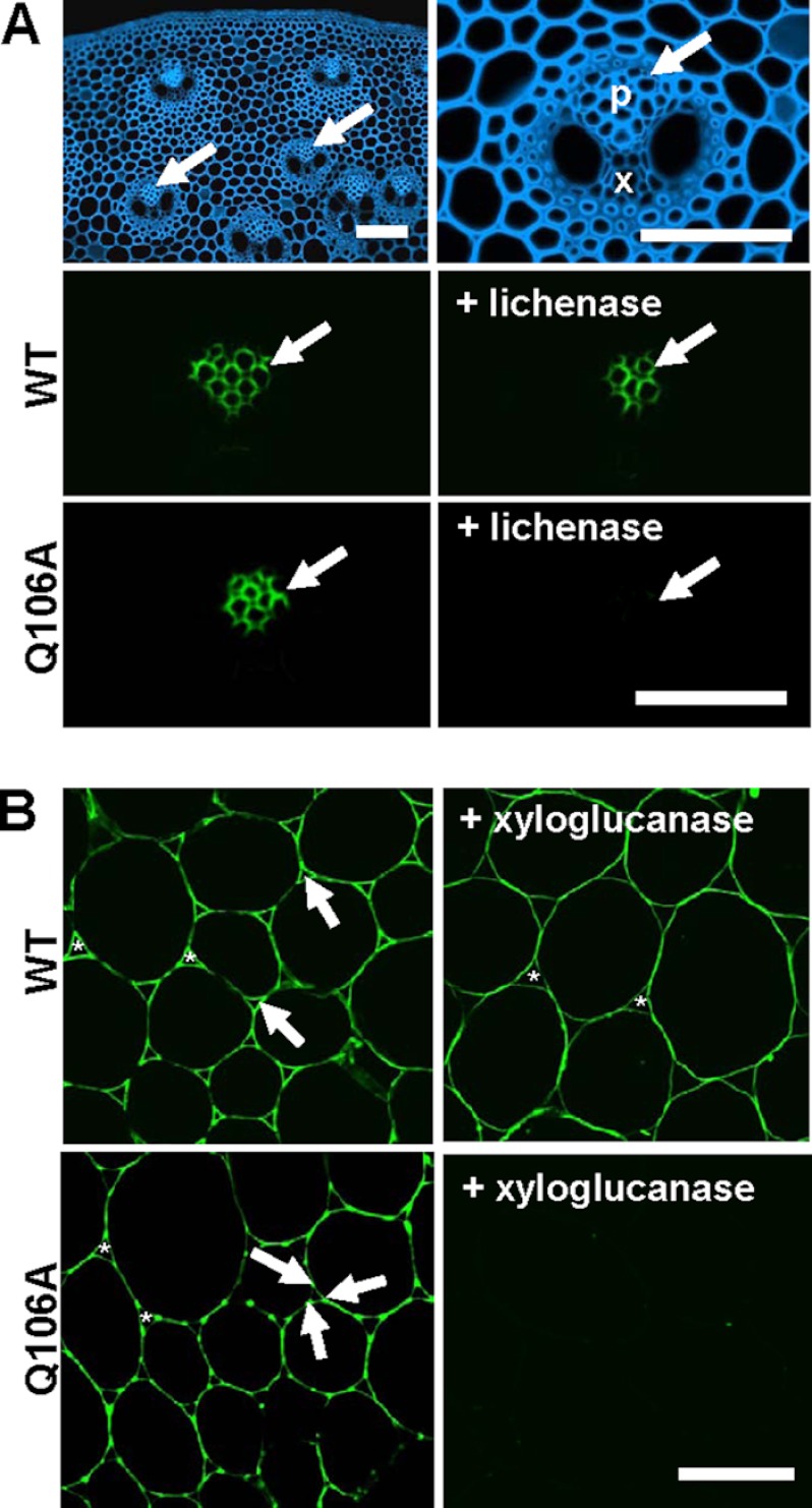
Plant biomass is central to the carbon cycle and to environmentally sustainable industries exemplified by the biofuel sector. Plant cell wall degrading enzymes generally contain noncatalytic carbohydrate binding modules (CBMs) that fulfil a targeting function, which enhances catalysis. CBMs that bind β-glucan chains often display broad specificity recognizing β1,4-glucans (cellulose), β1,3-β1,4-mixed linked glucans and xyloglucan, a β1,4-glucan decorated with α1,6-xylose residues, by targeting structures common to the three polysaccharides. Thus, CBMs that recognize xyloglucan target the β1,4-glucan backbone and only accommodate the xylose decorations. Here we show that two closely related CBMs, CBM65A and CBM65B, derived from EcCel5A, a Eubacterium cellulosolvens endoglucanase, bind to a range of β-glucans but, uniquely, display significant preference for xyloglucan. The structures of the two CBMs reveal a β-sandwich fold. The ligand binding site comprises the β-sheet that forms the concave surface of the proteins. Binding to the backbone chains of β-glucans is mediated primarily by five aromatic residues that also make hydrophobic interactions with the xylose side chains of xyloglucan, conferring the distinctive specificity of the CBMs for the decorated polysaccharide. Significantly, and in contrast to other CBMs that recognize β-glucans, CBM65A utilizes different polar residues to bind cellulose and mixed linked glucans. Thus, Gln(106) is central to cellulose recognition, but is not required for binding to mixed linked glucans. This report reveals the mechanism by which β-glucan-specific CBMs can distinguish between linear and mixed linked glucans, and show how these CBMs can exploit an extensive hydrophobic platform to target the side chains of decorated β-glucans.


Figures

FIGURE 1.
FIGURE 1.
Schematic of EcCel5A.
FIGURE 2.
FIGURE 2.
Examples of affinity gel electrophoresis of CBM65A and CBM65B against soluble polysaccharides. Panel A, the two CBM65 proteins were electrophoresed on nondenaturating polyacrylamide gels containing no ligand (control) or 0.3 mg/ml of the target polysaccharide (HEC, hydroxyethylcellulose). BSA was used as a nonpolysaccharide binding control. Panel B, wild type (WT) and mutants of CBM65A were electrophoresed in the presence or absence of the stated polysaccharides.
FIGURE 3.
FIGURE 3.
Representative ITC data of CtCBM62 binding to soluble ligands. The ligand (C6, cellohexaose; XG, xyloglucan; β-Glu, barley β-glucan) in the syringe was titrated into CBM65A or Q106A (100 μm) in the cell. The top half of each panel shows the raw ITC heats; the bottom half shows the integrated peak areas fitted using one single binding model by MicroCal Origin software. ITC was carried out in 50 mm Na-HEPES, pH 7.5, at 25 °C.
FIGURE 4.
FIGURE 4.
Structure of CBM65A. Panel A depicts CBM65A and CBM65B as a protein schematic, continuously color ramped from N to C terminus, from blue to red. The ligand binding residues are drawn as sticks. The arrows point to the loop in CBM65A and CBM65B that contain cellohexaose binding residues Gln106 and Asp649, respectively. Panel B shows a stereo representation of the ligand electron density (2FoFc) at 1.5 σ. CBM65B is shown as a schematic representation colored as in panel A. XXXG is shown in stick format with Glc and Xyl carbons colored yellow and magenta, respectively. Panel C shows the solvent-accessible surface of CBM65B with XXXG bound to the surface with the ligand binding aromatic residues shown in green. Panel D shows an overlay of the ligand binding site of CBM65A (carbons of amino acids shown in green) and CBM65B (carbons of amino acids shown in cyan). Dashed lines between atoms show hydrogen bonds. The figure was drawn with PyMOL.
FIGURE 5.
FIGURE 5.
Immunofluorescence analysis of CBM65A binding to cell walls in situ. Panel A, transverse sections of M. x giganteus stem. Calcofluor white shows staining of all cell walls (blue) and anatomy of a vascular bundle. In untreated sections both CBMs bind specifically to cell walls of the phloem (p) regions indicated by arrows; x = xylem. After lichenase pre-treatment of the section, before immunofluorescence analysis, wild type CBM65A (WT) still binds to the phloem cell walls but Q106A does not. All fluorescence micrographs have equivalent exposure times. Panel B, transverse sections of tobacco stem showing cell walls in the region of the pith parenchyma after pre-treatment with pectate lyase to remove pectic homogalacturonan. WT and Q106A displayed differential binding to parenchyma cell walls with WT binding strongly to cell walls and particularly to cell wall regions lining intercellular spaces (*) as indicated by arrows (exposure time 25 ms). Q106A bound less strongly to cell walls (exposure time 200 ms) and displayed some preferential binding to adhered cell wall regions at the corners of intercellular spaces; xyloglucan is known to be preferentially located in these regions (23). After a section pre-treatment with a xyloglucan-specific xyloglucanase, WT bound evenly to all cell walls with no differential binding in relationship to intercellular spaces, whereas Q106A did not bind (exposure time for both +xyloglucanase micrographs, 600 ms). Scale bars = 100 μm.

References

    1. Flint H. J., Duncan S. H., Scott K. P., Louis P. (2007) Interactions and competition within the microbial community of the human colon. Links between diet and health. Environ. Microbiol. 9, 1101–1111 - PubMed
    1. Mackie R. I., White B. A. (1990) Recent advances in rumen microbial ecology and metabolism. Potential impact on nutrient output. J. Dairy Sci. 73, 2971–2995 - PubMed
    1. Himmel M. E., Bayer E. A. (2009) Lignocellulose conversion to biofuels. Current challenges, global perspectives. Curr. Opin. Biotechnol. 20, 316–317 - PubMed
    1. Himmel M. E., Ding S. Y., Johnson D. K., Adney W. S., Nimlos M. R., Brady J. W., Foust T. D. (2007) Biomass recalcitrance. Engineering plants and enzymes for biofuels production. Science 315, 804–807 - PubMed
    1. Gilbert H. J. (2010) The biochemistry and structural biology of plant cell wall deconstruction. Plant Physiol. 153, 444–455 - PMC - PubMed

Publication types

MeSH terms