Review
. 2013 Jan;14(1):39-48.
doi: 10.1038/embor.2012.197. Epub 2012 Dec 11.

A matter of life and death: self-renewal in stem cells

Affiliations
Review

A matter of life and death: self-renewal in stem cells

Elaine Fuchs et al. EMBO Rep. 2013 Jan.

Abstract

If Narcissus could have self-renewed even once on seeing his own reflection, he would have died a happy man. Stem cells, on the other hand, have an enormous capacity for self-renewal; in other words, the ability to replicate and generate more of the same. In adult organisms, stem cells reside in specialized niches within each tissue. They replenish tissue cells that are lost during normal homeostasis, and on injury they repair damaged tissue. The ability of a stem cell to self-renew is governed by the dynamic interaction between the intrinsic proteins it expresses and the extrinsic signals that it receives from the niche microenvironment. Understanding the mechanisms governing when to proliferate and when to differentiate is vital, not only to normal stem cell biology, but also to ageing and cancer. This review focuses on elucidating conceptually, experimentally and mechanistically, our understanding of adult stem cell self-renewal. We use skin as a paradigm for discussing many of the salient points about this process, but also draw on the knowledge gained from these and other adult stem cell systems to delineate shared underlying principles, as well as highlight mechanistic distinctions among adult tissue stem cells. By doing so, we pinpoint important questions that still await answers.


Conflict of interest statement

The authors declare that they have no conflict of interest.

Figures

Figure 1
Figure 1
Timing and frequency of adult stem cell self-renewal proliferation. (A) Self-renewal of mouse HFSCs. Hair follicles undergo cycles of growth (pink box), degeneration (not shown) and rest (blue box). HFSCs are located in a niche called ‘the bulge’, in which they normally exist in a quiescent state, as represented by green cells. They only become activated just after the growth phase has begun, where they self-renew (as indicated by red cells) for a few days before returning to quiescence. HFSCs that exit the bulge niche proliferate earlier and remain proliferative slightly longer than those in the bulge. This leads to generation of more HFSCs that form a new bulge as the degenerative phase ends and the follicle returns to rest. (B) Self-renewal proliferation of mouse ISCs. Intestine epithelium constantly regenerates. It takes approximately 3–5 days for cells near the base of the crypt to be shed from the villus tip. ISCs reside at the bottom of the crypt, where they self-renew continuously, dividing once per day. (C) Activation of mouse HSCs. Most HSCs reside in the bone marrow, in two separate niches: the endosteum and central marrow. The most quiescent HSCs retain nucleotide and/or fluorescent histone labelling and are sometimes referred to as LRCs (see text). They divide approximately once every 4–5 months during normal homeostasis. Although not definitively shown, some researchers have proposed that the more active HSCs divide approximately once a month and tend to localize in the central marrow, close to the vasculature, where they enter and circulate in the bloodstream. HFSC, hair follicle stem cell; HSC, haematopoietic stem cell; ISC, intestine stem cell; LRC, label retaining cell; SC, stem cell.
Figure 2
Figure 2
Balancing stem cell self-renewal and tissue regeneration. (A, A') Molecular and lineage-tracing studies have demonstrated that HFSCs (green, quiescent; red, proliferative) support follicle homeostasis and hair growth and, at the same time, maintain the pool of undifferentiated stem cells. Activation, proliferation and fate commitment of HFSCs are achieved in part through Wnt signalling and BMP inhibition (A), which reaches a crucial threshold at the transition that mobilizes the stem cell niche to make a new hair follicle (A). Activated HFSCs maintaining contact with the dermal papilla (brown) soon commit to form transit-amplifying (TA) matrix progenitors (blue), and can never again be stem cells. As the follicle grows downwards and matures, BMP inhibition and Wnt signalling at the base (hair bulb) continue to increase, and matrix cells rapidly divide several times, then terminally differentiate to produce the hair and the inner cells of the hair follicle (A'). At the end of the growth phase, HFSCs that exited the niche to the upper portion of the outer root sheath (ORS) return to form the new bulge and hair germ. Those further along in transit between the bulge and the matrix reach a cell-fate commitment ‘point of no return’. However, they too move back to the niche, where they become the keratin 6-positive (K6+) inner bulge cells (shown in orange), which further suppress HFSC self-renewal by expressing inhibitory signals such as BMPs [5]. (B) Lineage-tracing experiments have demonstrated that ISCs (shown in green) self-renew long term and generate all of the differentiated progeny in the intestinal epithelium, including Paneth cells (shown in orange), which maintain self-renewal within the stem cell niche in part by expressing Wnt signals [39]. (C) Downstream HSC progenies participate in regulating HSC activation. HSCs first generate MPPs that are similar in concept to the intermediate, unspecified proliferative matrix cells of the hair follicle. MPPs then differentiate into one of several haematopoietic lineages. Macrophages are one of the terminally differentiated cell types of the HSC lineage [15]. They circulate and return to the niche, where they provide inhibitory signals that restrict the number of HSCs that become activated for circulation. Within the bone marrow, the niche for HSCs was initially thought to be the osteoblast lining of the bone, but evidence points to a more important role for perivascular and endothelial cells in HSC maintenance [94]. BMP, bone morphogenetic protein; HFSC, hair follicle stem cell; HSC, haematopoietic stem cell; ISC, intestinal stem cell; MPP, multipotent progenitor; Wnt, mammalian homologue of Drosophila ‘wingless’ signalling protein.
Figure 3
Figure 3
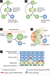
Symmetrical as opposed to asymmetrical cell division to achieve self-renewal and differentiation. (A) In asymmetrical cell divisions, cell fate specification is coupled to mitosis and involves unequal partitioning of cellular components to the resulting daughter cells. (B) In symmetrical cell divisions, cellular components are equally distributed to the two daughter cells. Although cell fate specification is not coupled to mitosis in a symmetrical division, it can be intimately linked if one of the two daughter cells is displaced to a new microenvironment. (C) Drosophila neuroblasts divide by asymmetrical cell division. To partition cellular components unequally, they use a polarized Par3/aPKC-containing apical crescent (marked in red) to establish cell polarity. As the neuroblast enters mitosis, the apical crescent recruits additional proteins that polarize the spindle and differentially partition Notch signalling components, such that on completion of mitosis, the cell retaining the apical crescent remains as a stem cell and the other becomes the differentiated GMC. (D) Drosophila male GSCs also divide asymmetrically, but through a different mechanism. GSCs surround the Hub cells located at the tip of the testis. They are interspersed with somatic CySCs. GSCs attach to the niche Hub cells through adherens junctions (marked in purple), which polarize the mother centriole (marked in red) and align the GSC spindle in a fashion such that one daughter retains the mother centriole and its association with the Hub, whilst the other becomes fated to differentiate as a gonialblast. (E) Mouse embryonic basal epidermal cells undergo asymmetrical cell divisions. These cells are polarized through intercellular adherens junctions (marked in purple), and basement membrane–substratum junctions (marked in blue). These features localize an apical crescent, which serves as a platform for recruiting some of the same spindle-orienting, asymmetrical division components as the fly neuroblast. After divisions perpendicular to the basement membrane, one daughter probably inherits integrins and associated growth factor receptors disproportionately, remaining as a basal stem cell, whereas the displaced suprabasal daughter cell differentially inherits Notch signalling. Approximately 60% of embryonic basal epidermal cells undergo asymmetrical division whilst the remaining ones undergo symmetrical cell division relative to the basement membrane. aPKC, atypical protein kinase C; CySC, cyst stem cell; GMC, ganglion mother cell; GSC, germline stem cell; Par3, partitioning defective protein 3; SC, stem cell.
Figure 4
Figure 4
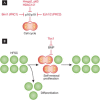
Intrinsic factors regulating stem cell self-renewal. (A) p16Ink4a is a potent inhibitor of the G1- to S-phase transition in the cell cycle. To allow cell cycle entry, p16Ink4a must be repressed. Many factors have been reported to silence p16Ink4a gene expression in adult stem cells including Bmi1, Hmga2, p63, Ezh1/2 and HDAC1/2. (B) Some self-renewal factors seem to be tailored to suit the context-dependent needs of a stem cell niche. One example is HFSC-enriched factor Tbx1, which suppresses BMP signalling to allow HFSC self-renewal in the BMP-high microenvironment of the bulge niche. Bmi1, B-lymphoma Moloney murine leukaemia virus insertion region 1; BMP, bone morphogenetic protein; Ezh1/2, enhancer of zeste homologue 1/2; HDAC1/2, histone deacetylase 1/2; HFSC, hair follicle stem cell; Hmga2, high mobility group AT-hook 2; p16Ink4a, tumour suppressor protein encoded by the Ink4a gene locus; Tbx1, T-box transcription factor 1.
None
Elaine Fuchs
None
Ting Chen

References

    1. Copley MR, Beer PA, Eaves CJ (2012) Hematopoietic stem cell heterogeneity takes center stage. Cell Stem Cell 10: 690–697 - PubMed
    1. Barker N, van Oudenaarden A, Clevers H (2012) Identifying the stem cell of the intestinal crypt: strategies and pitfalls. Cell Stem Cell 11: 452–460 - PubMed
    1. He S, Nakada D, Morrison SJ (2009) Mechanisms of stem cell self-renewal. Annu Rev Cell Dev Biol 25: 377–406 - PubMed
    1. O'Brien LE et al. (2011) Altered modes of stem cell division drive adaptive intestinal growth. Cell 147: 603–614 - PMC - PubMed
    1. Hsu YC, Pasolli HA, Fuchs E (2011) Dynamics between stem cells, niche, and progeny in the hair follicle. Cell 144: 92–105 - PMC - PubMed

Publication types