. 2012 Dec 10;199(6):883-91.
doi: 10.1083/jcb.201208024.

Munc13 controls the location and efficiency of dense-core vesicle release in neurons

Affiliations

Munc13 controls the location and efficiency of dense-core vesicle release in neurons

Rhea van de Bospoort et al. J Cell Biol. .

Abstract

Neuronal dense-core vesicles (DCVs) contain diverse cargo crucial for brain development and function, but the mechanisms that control their release are largely unknown. We quantified activity-dependent DCV release in hippocampal neurons at single vesicle resolution. DCVs fused preferentially at synaptic terminals. DCVs also fused at extrasynaptic sites but only after prolonged stimulation. In munc13-1/2-null mutant neurons, synaptic DCV release was reduced but not abolished, and synaptic preference was lost. The remaining fusion required prolonged stimulation, similar to extrasynaptic fusion in wild-type neurons. Conversely, Munc13-1 overexpression (M13OE) promoted extrasynaptic DCV release, also without prolonged stimulation. Thus, Munc13-1/2 facilitate DCV fusion but, unlike for synaptic vesicles, are not essential for DCV release, and M13OE is sufficient to produce efficient DCV release extrasynaptically.


Figures
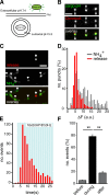
Figure 1.
Figure 1.
Optical sensor for DCV release in neurons. (A) Schematic representation of an optical reporter for DCV release that allows visualization of single DCV fusion events. PM, plasma membrane. (B) Confocal image of a neuron transfected with Semaphorin-3a–pHluorin (cargo-pH) and stained for endogenous DCV cargo chromogranin A (ChromoA) showing complete overlap (arrowheads). (C) Image series showing a cargo-pHluorin release event (arrows) and the NH4+ response to reveal all vesicles in the neurite (arrowheads). (D) Normalized frequency distribution of ΔF for DCV release events measured with Semaphorin-3a–pHluorin and ΔF upon NH4+ perfusion (NH4+: 395 puncta, 7 cells; release: 293 puncta, 30 cells; median ΔF release = 5.4 a.u.; median ΔF NH4+ = 6.0 a.u.). (E) Frequency distribution of DCV release events measured with Semaphorin-3a–pHluorin (570 release events in 53 cells; blue bars, 16 bursts of 50 APs at 50 Hz). (F) DCV release events during 60 s before stimulation, during stimulation, and during 70 s after stimulation (before: 1 ± 0.6; during: 16 ± 3.4; after: 5 ± 1.3 vesicles/cell; n = 21 cells, n = 3). **, P < 0.01. Data are plotted as means with SEM. Bars, 2 µm.
Figure 2.
Figure 2.
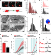
DCV release is enriched and more efficient at synapses although DCVs do not accumulate at synaptic regions. (A) Example of a synaptic (filled arrowheads) and an extrasynaptic release event (open arrowheads). Synapsin-mCherry is shown in red, and release events are shown in green. Bar, 1 µm. (B) Cartoon showing a synaptic (left green dot) and extrasynaptic (right green dot) release event. (C) Overlay of frequency distribution of ΔF upon NH4+ application for synaptic (syn) DCV puncta (synaptic 589 puncta) and extrasynaptic (e-syn) DCV puncta (extrasynaptic 612 puncta). (D) Individual frequency distributions of ΔF upon NH4+ application. (E) Electron micrographs of synaptic terminals that harbor DCVs. Arrowheads point to DCVs. Bars, 100 nm. (F) Diameter of DCVs in electron micrographs (mean diameter: 68.9 ± 1.0 nm, 146 DCVs in 7 cells). (G) Number of DCVs per synaptic profile (102 synapses in 7 cells). Inset, ±22% of synaptic DCVs is docked to the plasma membrane (undocked, 113; docked, 33; total, 146). (H) Percentage of synaptic and extrasynaptic DCV release events measured with Semaphorin-3a–pHluorin (235 synaptic release events, 131 extrasynaptic release events; n = 34 cells, n = 3; *, P < 0.05). (I) Cumulative number of release events per cell for synaptic and extrasynaptic events. (Synaptic release rate: 0.26 ± 0.03 vesicles/s; extrasynaptic: 0.13 ± 0.02 vesicles/s). Shading represents SEM. (J) Cumulative number of events during the first four bursts (blue bars 1, 2, 3, and 4 represent 50 APs at 50 Hz) for synaptic and extrasynaptic events. (K) Example traces of synaptic (red) and extrasynaptic (black) release events. Arrows mark start of the stimulation. Data are plotted as means with SEM.
Figure 3.
Figure 3.
Munc13 specifically reduces DCV release at synapses. (A) Mean number of DCV release events per cell for WT and Munc13-1/2 DKO (M13DKO) neurons measured with Semaphorin-3a–pHluorin (WT: 482 events, n = 41 cells; M13DKO: 108 events, n = 23 cells; n = 4; **, P < 0.01). (B) Cumulative number of release events per cell. (C) Cumulative (cum.) number of release events during the first four bursts of 50 APs at 50 Hz. (D) Percentage of synaptically localized DCVs that remain synaptic during 10-s acquisition (WT: 221 vesicles, n = 6 cells; M13DKO: 146 vesicles, n = 5 cells; n = 2). (E) Percentage of synaptic and extrasynaptic DCV release events in WT and M13DKO neurons (WT: 21 cells; M13DKO: 23 cells; n = 4; *, P < 0.05; **, P < 0.01). (F) Total numbers of synaptic and extrasynaptic events show a specific loss of synaptic events (total events: WT, 255; M13DKO, 151). Red dotted line shows difference in bar heights. (G) Cumulative number of synaptic DCV release events. (H) Cumulative number of extrasynaptic DCV release events. Shading represents SEM. Data are plotted as means with SEM.
Figure 4.
Figure 4.
M13OE increases DCV release at extrasynaptic sites. (A) Normalized Munc13-1 intensity in WT neurons compared with neurons overexpressing Munc13-1 (M13OE, n = 3; **, P < 0.001). (B) Confocal image of a WT neuron with endogenous Munc13-1 and a WT neuron overexpressing Munc13-1 (arrowheads show synaptic localization of Munc13). Bars, 2 µm. (C) Percentage of synaptically localized DCVs that stay synaptic for 10 s (WT: 199 vesicles, n = 7 cells; M13OE: 170 vesicles, n = 7 cells). (D) Mean number of DCV release events per cell (WT: 361 events in 36 cells; M13OE: 588 events in 32 cells; n = 3; *, P < 0.05). (E) Cumulative (cum.) DCV release events per cell. (F) Cumulative number of release events during the first four bursts of 50 APs at 50 Hz. (G) Percentage of synaptic and extrasynaptic DCV release events (**, P < 0.01; *, P < 0.05). (H) Total numbers of synaptic and extrasynaptic DCV release events. Loss of synaptic preference is caused by an increase of extrasynaptic release events in M13OE. Red dotted line shows difference in bar heights. (I) Cumulative extrasynaptic DCV release events. (J) Cumulative number of events during the first four bursts at extrasynaptic sites. (K) Cumulative synaptic DCV release events. (L) Cumulative number of events during the first four bursts at synapses. Shading represents SEM. Data are plotted as means with SEM.

References

    1. Ashery U., Varoqueaux F., Voets T., Betz A., Thakur P., Koch H., Neher E., Brose N., Rettig J. 2000. Munc13-1 acts as a priming factor for large dense-core vesicles in bovine chromaffin cells. EMBO J. 19:3586–3596 10.1093/emboj/19.14.3586 - DOI - PMC - PubMed
    1. Augustin I., Rosenmund C., Südhof T.C., Brose N. 1999. Munc13-1 is essential for fusion competence of glutamatergic synaptic vesicles. Nature. 400:457–461 10.1038/22768 - DOI - PubMed
    1. Bartfai T., Iverfeldt K., Fisone G., Serfözö P. 1988. Regulation of the release of coexisting neurotransmitters. Annu. Rev. Pharmacol. Toxicol. 28:285–310 10.1146/annurev.pa.28.040188.001441 - DOI - PubMed
    1. Basu J., Betz A., Brose N., Rosenmund C. 2007. Munc13-1 C1 domain activation lowers the energy barrier for synaptic vesicle fusion. J. Neurosci. 27:1200–1210 10.1523/JNEUROSCI.4908-06.2007 - DOI - PMC - PubMed
    1. Deng L., Kaeser P.S., Xu W., Südhof T.C. 2011. RIM proteins activate vesicle priming by reversing autoinhibitory homodimerization of Munc13. Neuron. 69:317–331 10.1016/j.neuron.2011.01.005 - DOI - PMC - PubMed

Publication types

MeSH terms

Substances