Review
. 2013 Aug;54(8):2034-2048.
doi: 10.1194/jlr.R034025. Epub 2012 Dec 10.

New insights into the determination of HDL structure by apolipoproteins: Thematic review series: high density lipoprotein structure, function, and metabolism

Affiliations
Review

New insights into the determination of HDL structure by apolipoproteins: Thematic review series: high density lipoprotein structure, function, and metabolism

Michael C Phillips. J Lipid Res. 2013 Aug.

Abstract

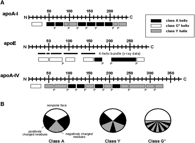
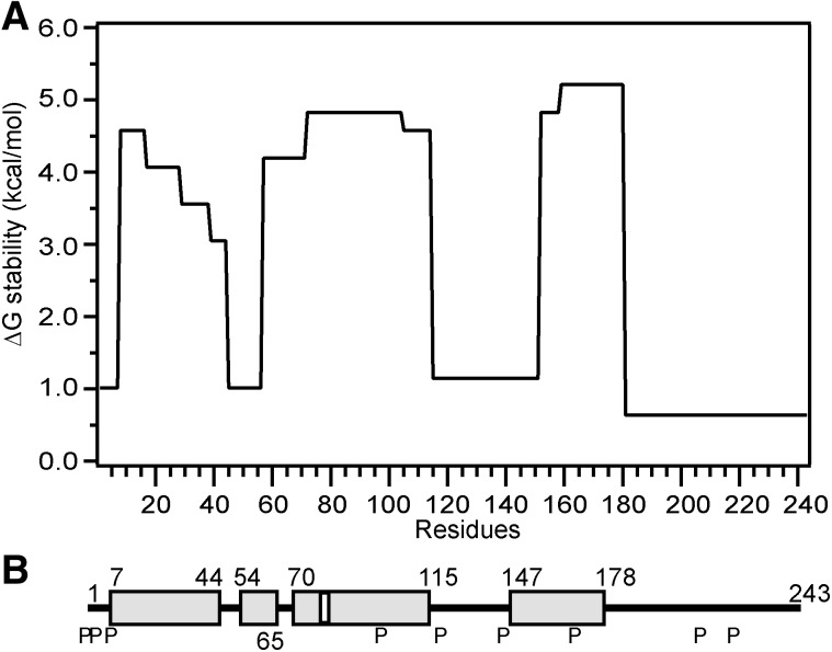
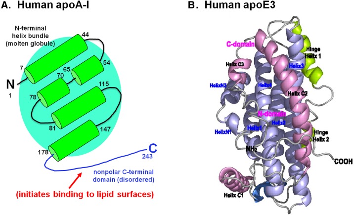
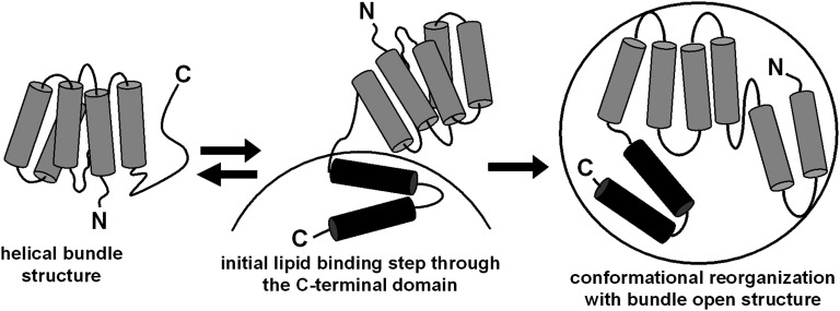
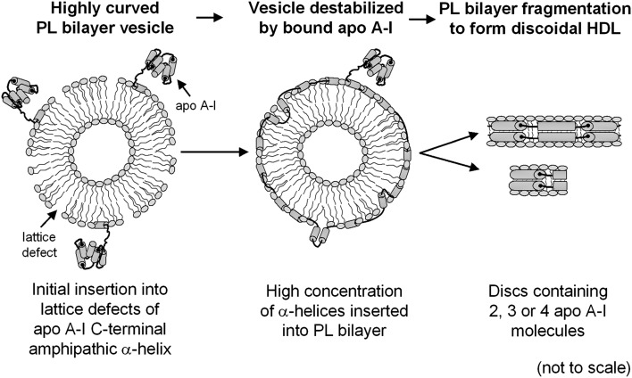
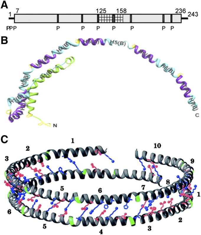
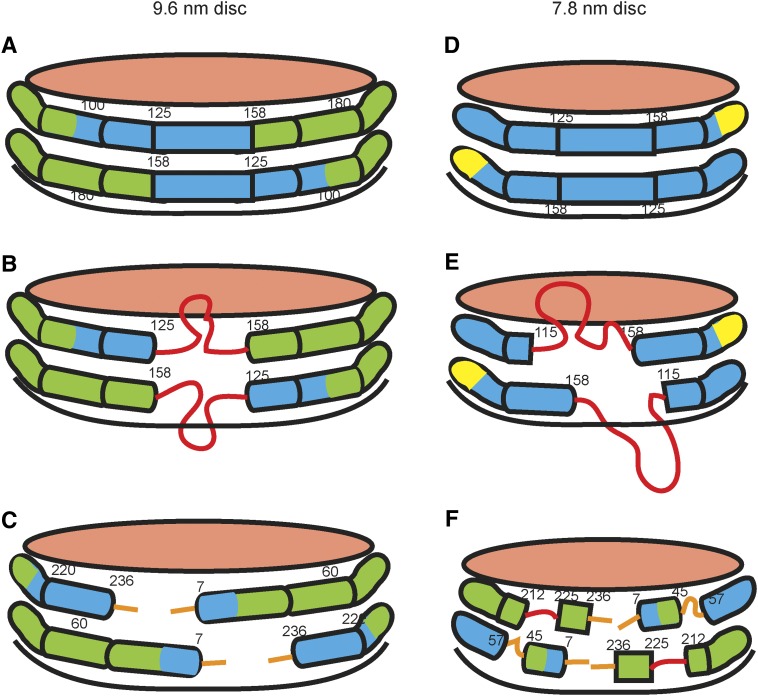
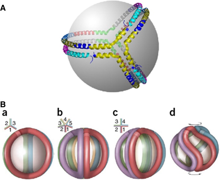
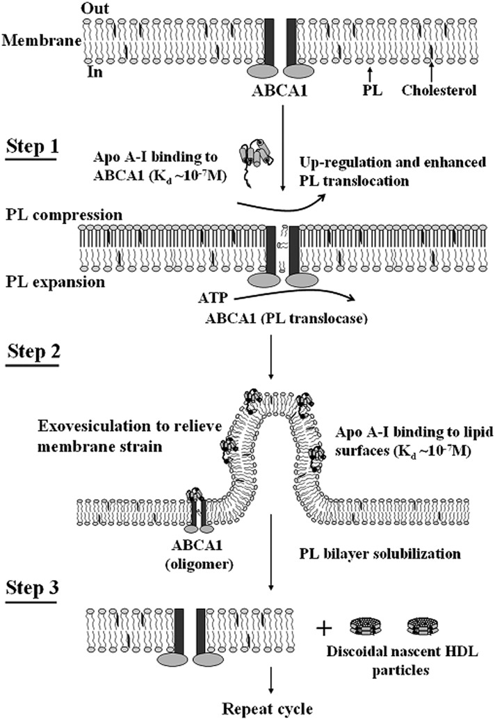
Apolipoprotein (apo)A-I is the principal protein component of HDL, and because of its conformational adaptability, it can stabilize all HDL subclasses. The amphipathic α-helix is the structural motif that enables apoA-I to achieve this functionality. In the lipid-free state, the helical segments unfold and refold in seconds and are located in the N-terminal two thirds of the molecule where they are loosely packed as a dynamic, four-helix bundle. The C-terminal third of the protein forms an intrinsically disordered domain that mediates initial binding to phospholipid surfaces, which occurs with coupled α-helix formation. The lipid affinity of apoA-I confers detergent-like properties; it can solubilize vesicular phospholipids to create discoidal HDL particles with diameters of approximately 10 nm. Such particles contain a segment of phospholipid bilayer and are stabilized by two apoA-I molecules that are arranged in an anti-parallel, double-belt conformation around the edge of the disc, shielding the hydrophobic phospholipid acyl chains from exposure to water. The apoA-I molecules are in a highly dynamic state, and they stabilize discoidal particles of different sizes by certain segments forming loops that detach reversibly from the particle surface. The flexible apoA-I molecule adapts to the surface of spherical HDL particles by bending and forming a stabilizing trefoil scaffold structure. The above characteristics of apoA-I enable it to partner with ABCA1 in mediating efflux of cellular phospholipid and cholesterol and formation of a heterogeneous population of nascent HDL particles. Novel insights into the structure-function relationships of apoA-I should help reveal mechanisms by which HDL subclass distribution can be manipulated.

Keywords: ATP binding cassette transporter A1; amphipathic α-helix; apoA-I; apoE; cholesterol; helix bundle; lipoprotein; membrane solubilization; phospholipid.


Figures

Fig. 1.
Fig. 1.
(A) Distribution of amphipathic α-helices in the human exchangeable apolipoproteins apoA-I, apoA-IV, and apoE. The letter “P” below the rectangles indicates positions of all proline residues. (B) Amphipathic helix classes found in the exchangeable apolipoproteins. Classification is based on the distribution of charged residues. Adapted from Segrest et al. (12).
Fig. 2.
Fig. 2.
Summary of the HX-derived secondary structure assignments and α-helix stabilities for lipid-free human apoA-I. (A) Site-resolved stability of apoA-I in the monomeric state. (B) The gray cylinders represent α-helices, and the lines indicate disordered secondary structure. The positions of proline residues (P), whose presence leads to some perturbation of α-helix organization, are marked. The helical structure is dynamic, unfolding and refolding in seconds. Adapted from Ref. .
Fig. 3.
Fig. 3.
Structures of human apoA-I and apoE3 in the lipid-free monomeric state. Both molecules adopt a two-domain tertiary structure in which the N-terminal two thirds is folded into an anti-parallel helix bundle, and the C-terminal region forms a distinct domain. (A) The helix locations along the amino acid sequence of the apoA-I molecule are taken from Fig. 2. (B) NMR structure of an apoE3 monomeric variant containing the mutations F257A/W264R/V269A/L279Q/V287E to prevent self-association. The average structure is shown as a ribbon representation in which the N-terminal helices in the anti-parallel bundle are colored blue. The C-terminal helices are shown in pink, and the hinge region that links the N- and C-terminal domains is colored green. ApoE3 figure reproduced with permission from Ref. .
Fig. 4.
Fig. 4.
Model of the two-step mechanism of human apoA-I binding to a spherical lipid particle. In the lipid-free state, apoA-I is organized into two structural domains as shown in Fig. 3A. Lipid binding is initiated by the C-terminal domain and is coupled to an increase in α-helicity. Subsequently, the helix bundle undergoes a conformational opening, converting hydrophobic helix-helix interactions to helix-lipid interactions; this second step is only slowly reversible. Reproduced with permission from Ref. .
Fig. 5.
Fig. 5.
Suggested molecular mechanism for the solubilization of PL bilayers by apoA-I to create discoidal HDL particles. Binding of apoA-I to the PL bilayer occurs in two steps as summarized in Fig. 4; formation of the C-terminal amphipathic α-helix by the apoA-I molecule is coupled with interaction of the protein with the PL surface. The discoidal HDL products of the solubilization process have structures like those depicted in Figs. 6C and 7. This process is envisaged to underlie the formation of nascent HDL particles in the apoA-I/ABCA1 reaction depicted in Fig. 9.
Fig. 6.
Fig. 6.
Structure of apoA-I in a discoidal HDL particle. (A) Summary of the HX-derived apoA-I helix locations in a 9.6 nm diameter particle that contains 140 molecules of POPC and 2 molecules of apoA-I. Residues 125–158 (cross-hatched) exist in two populations (helix and loop). The equivalent secondary structure information for lipid-free apoA-I is shown in the same representation in Fig. 2B. Reproduced with permission from Ref. . (B) Structure of apoA-I (Δ185–243) monomer obtained from a crystal structure at 2.2 Å resolution. This truncated apoA-I molecule adopts an extended conformation in the crystal and forms a half circle dimer. Reproduced with permission from Ref. . (C) Double-belt model for apoA-I structure at the edge of a discoidal HDL particle. Two ring-shaped molecules of apoA-I are stacked on top of each other with both molecules in an anti-parallel orientation, allowing the helix registry to maximize intermolecular salt-bridge interactions. Only the charged residues at selected positions are explicitly displayed; positively charged residues are represented in blue, negatively charged residues in red, and prolines in green. The numbers 1–10 represent the ten helical domains identified by computer analysis of the human apoA-I amino acid sequence between residues 44 and 243 (cf. Fig. 1A). Reproduced with permission from Ref. .
Fig. 7.
Fig. 7.
Diagram comparing the secondary structures of apoA-I molecules in 9.6 nm (A–C) and 7.8 nm (D–F) discoidal HDL particles, as determined by HX-MS. The amphipathic α-helices are represented by cylinders colored according to their stability. The values of the free energy of stabilization (kcal/mol) are as follows: red, ≤1; orange, 1–2; yellow, 2–3; green, 3–4; blue >4. Importantly, the structure is not static but highly dynamic; the α-helices unfold and refold in seconds or less. The pink surfaces of the discs are covered by PL polar groups. The apoA-I molecules are organized according to the double-belt model (Fig. 6C). (A) and (B) show the states in which residues 125–158 adopt α-helical and disordered loop conformations, respectively. The diagrams show the two apoA-I molecules on each disc acting in concert and adopting the same conformation, but the segment spanning residues 125–158 also could be α-helical in one molecule and loop in the other. The orientation in (C) is obtained by a 180° clockwise rotation of the disc in (A) and depicts the nonhelical structures formed by the 6 or 7 amino acids at the ends of the apoA-I molecules. (D–F) show the equivalent information for a 7.8 nm discoidal HDL particle. The approximately 20% smaller area available at the edge of the 7.8 nm disc leads to displacement of about 20% more apoA-I amino acid residues from contact with PL molecules and enhances formation of protruding disordered loops [red in (E) and (F)]. The surface-dissociated and surface-associated states coexist on the time scale of minutes or longer. Reproduced with permission from Ref. .
Fig. 8.
Fig. 8.
Models of apoA-I organization on spherical HDL particles. (A) Trefoil organization of three apoA-I molecules on the surface of a 9.6 nm diameter spherical particle. Each putative helical domain shown in Fig. 6C is represented as a separate color: helix 1, teal; helix 2, purple; helix 3, dark blue; helix 4, gray; helix 5, green; helix 6, red; helix 7, light blue; helix 8, dark yellow; helix 9, navy blue; and helix 10, yellow. All helix-to-helix interactions present in the double belt between two molecules of apoA-I in a disc (Fig. 6C) are also present among three apoA-I molecules in the trefoil. Reproduced with permission from Ref. . (B) Incorporation of additional apoA-I molecules to the trefoil model and apoA-I adaptation to smaller particle diameters. (a) Schematic representation of the three-molecule trefoil model with each molecule of apoA-I shown in a different color; See (A) for more detail. The lighter colored band on each molecule represents the N-terminus (residue 44, as the model was built in the absence of residues 1–43). The inset is a schematic top view showing the bend angles of each apoA-I. (b) Pentameric complex proposed for the structure of larger HDL. (c) An idealized, fully extended tetrameric complex. (d) Twisted tetrameric complex with a reduced particle diameter as proposed for smaller HDL. Reproduced with permission from Ref. (97).
Fig. 9.
Fig. 9.
Mechanism of interaction of apoA-I with ABCA1 and efflux of cellular phospholipids and cholesterol. The reaction in which apoA-I binds to ABCA1 and membrane lipids to create discoidal nascent HDL particles comprised three steps. Step 1 involves the high affinity binding of a small amount of apoA-I to ABCA1 located in the plasma membrane PL bilayer; this regulatory pool of apoA-I upregulates ABCA1 activity, thereby enhancing the active translocation of membrane PL from the cytoplasmic to exofacial leaflet. This translocase activity leads to lateral compression of the PL molecules in the exofacial leaflet and expansion of those in the cytoplasmic leaflet. Step 2 involves the bending of the membrane to relieve the strain induced by the unequal molecular packing density across the membrane and the formation of an exovesiculated domain to which apoA-I can bind with high affinity (cf. Fig. 4). This interaction with the highly curved membrane surface involves apoA-I/membrane lipid interactions and creates a relatively large pool of bound apoA-I. Step 3 involves the spontaneous solubilization (cf. Fig. 5) by the bound apoA-I of membrane PL and cholesterol in the exovesiculated domains to create discoidal HDL particles (cf. Figs. 6 and 7) containing two, three, or four apoA-I molecules/particle. In the diagram, the two transmembrane six-helix domains of ABCA1 are represented as rectangles, whereas the two ATPase domains are shown as ovals. The space between the two rectangles represents the chamber in which translocation of PL molecules occurs. Reproduced with permission from Ref. .

References

    1. Lund-Katz S., Liu L., Thuahnai S. T., Phillips M. C. 2003. High density lipoprotein structure. Front. Biosci. 8: d1044–d1054 - PubMed
    1. Kontush A., Chapman M. J. 2006. Functionally defective high-density lipoprotein: a new therapeutic target at the crossroads of dyslipidemia, inflammation, and atherosclerosis. Pharmacol. Rev. 58: 342–374 - PubMed
    1. Jonas A., Phillips M. C. 2008. Lipoprotein. In Biochemistry structure of Lipids, Lipoproteins and Membranes. 5th edition. D. E. Vance and J. E. Vance, editors. Elsevier, Oxford, UK. 485–506.
    1. Rothblat G. H., Phillips M. C. 2010. High-density lipoprotein heterogeneity and function in reverse cholesterol transport. Curr. Opin. Lipidol. 21: 229–238 - PMC - PubMed
    1. Shen B. W., Scanu A. M., Kezdy F. J. 1977. Structure of human serum lipoproteins inferred from compositional analysis. Proc. Natl. Acad. Sci. USA. 74: 837–841 - PMC - PubMed

Publication types