. 2012 Dec;2(12):1109-17.
doi: 10.1158/2159-8290.CD-12-0197.

Metabolomics strategy reveals subpopulation of liposarcomas sensitive to gemcitabine treatment

Affiliations

Metabolomics strategy reveals subpopulation of liposarcomas sensitive to gemcitabine treatment

Daniel Braas et al. Cancer Discov. 2012 Dec.

Abstract

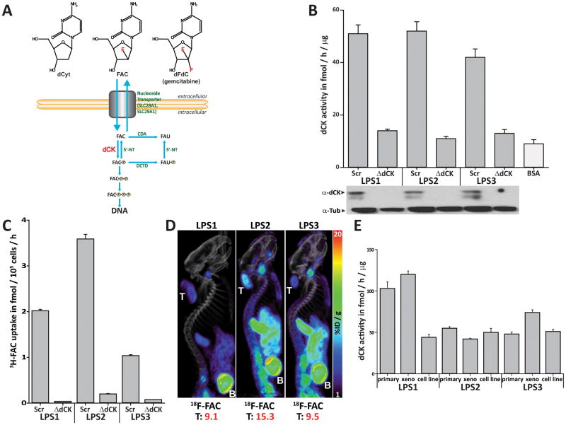
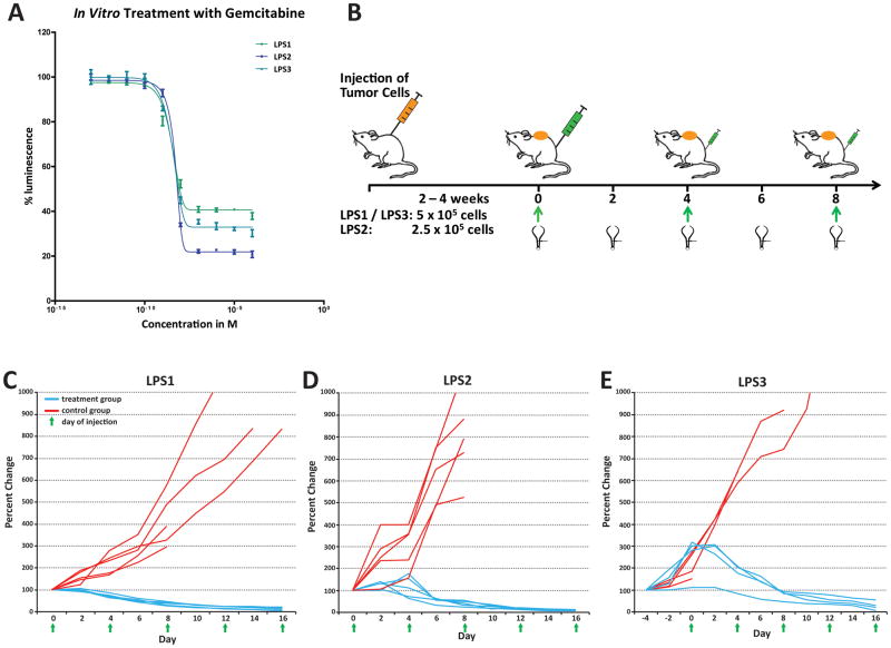
Unlike many cancers that exhibit glycolytic metabolism, high-grade liposarcomas often exhibit low 2[18F]fluoro-2-deoxy-D-glucose uptake by positron emission tomography (PET), despite rapid tumor growth. Here, we used liquid chromatography tandem mass spectrometry to identify carbon sources taken up by liposarcoma cell lines derived from xenograft tumors in patients. Interestingly, we found that liposarcoma cell lines consume nucleosides from culture media, suggesting nucleoside salvage pathway activity. The nucleoside salvage pathway is dependent on deoxycytidine kinase (dCK) and can be imaged in vivo by PET with 1-(2'-deoxy-2'-[18F]fluoroarabinofuranosyl) cytosine (FAC). We found that liposarcoma cell lines and xenograft tumors exhibit dCK activity and dCK-dependent FAC uptake in vitro and in vivo. In addition, liposarcoma cell lines and xenograft tumors are sensitive to treatment with the nucleoside analogue prodrug gemcitabine, and gemcitabine sensitivity is dependent on dCK expression. Elevated dCK activity is evident in 7 of 68 clinical liposarcoma samples analyzed. These data suggest that a subpopulation of liposarcoma patients have tumors with nucleoside salvage pathway activity that can be identified noninvasively using [18F]-FAC-PET and targeted using gemcitabine.

Significance: Patients with high-grade liposarcoma have poor prognoses and often fail to respond to chemotherapy. This report identifies elevated nucleoside salvage activity in a subset of liposarcomas that are identifiable using noninvasive PET imaging with FAC and that are sensitive to gemcitabine. Thus, we suggest a new treatment paradigm for liposarcoma patients that uses [18F]-FAC-PET in the clinic to delineate gemcitabine responders from nonresponders.


Conflict of interest statement

Conflict of interest statement: ONW is an inventor of national and Patent Cooperation Treaty (PCT) patent applications for FAC technology referred to in the article. ONW is also involved in Sofie Biosciences, a startup company that has licensed this intellectual property. All other authors declare no conflict of interest.

Figures

Figure 1
Figure 1. Mass spectrometry-based metabolomics identifies nucleosides amongst other nutrients consumed by liposarcoma cell lines in vitro
Metabolites were extracted from media samples from cultured LPS cells and analyzed using LC-MS/MS. A, Metabolomic footprint analysis for LPS2 cells is shown with spectral counts depicted as a heatmap. B, Venn-diagram depicting overlapping consumption of metabolites in liposarcoma cell lines (LPS1-3). The ten metabolites commonly consumed by the cell lines analyzed are listed below. C–E, Relative abundance of cytidine, thymidine and uridine in media samples plotted over time for each cell line studied.
Figure 2
Figure 2. Liposarcoma cell lines and xenograft tumors exhibit nucleoside salvage pathway activity
A, Schematic representation of the nucleoside salvage pathway. dCK (red) catalyzes the rate-limiting initial phosphorylation step in the nucleoside salvage pathway important for trapping deoxycytidine, deoxyguanosine, deoxyadenosine, and FAC inside the cell and activating gemcitabine. ForB–C, liposarcoma cell lines (LPS1-3) were engineered to stably express a scrambled shRNA (scr) or a shRNA construct towards dCK (ΔdCK), and dCK activity (B) and 3H-FAC uptake measurements (C) confirm nucleoside salvage activity in vitro. D, MicroPET/CT imaging of liposarcoma xenografts in NSG mice with [18F]-FAC indicates nucleoside salvage activity in vivo. E, Comparison of dCK activity in lysates from corresponding primary tumors, xenograft tumors, and cell lines. Xeno = xenograft tumor.
Figure 3
Figure 3. The nucleoside prodrug gemcitabine is cytotoxic to liposarcoma cell lines and xenograft tumors in mice
A, Gemcitabine treatment reduces viability of liposarcoma cell lines in vitro. B, Schematic representation of the liposarcoma xenograft tumor gemcitabine treatment experiment. Xenograft tumors were established by subcutaneous injection of 5×10⁵ LPS1 (C, n=10) and LPS3 (E, n=8) or 2.5×10⁵ LPS2 (D, n=10) cells. Tumors were measured every second day. Gemcitabine treatment was given by intraperitoneal injection every fourth day (green arrows). Mice in the control group were injected with the same volume of PBS. C–E, Gemcitabine treatment leads to regression of liposarcoma xenograft tumors in mice derived from three liposarcoma cell lines (LPS1-3).
Figure 4
Figure 4. dCK activity is elevated in 10% of liposarcomas and is required for response to gemcitabine treatment
A, dCK activity is elevated in 7 out of 68 (10%) primary liposarcoma tumor samples. Protein extracts were prepared from 68 primary liposarcoma (including LPS 1–3, green) tumors, and dCK activity was compared to BSA, WT and dCK−/− mouse splenocytes (orange). Dotted line indicates average dCK activity across measured samples. B, Gemcitabine sensitivity is dependent on dCK expression in vitro. Stable knockdown of dCK (ΔdCK) in LPS2 cells increases the LC50 of gemcitabine relative to scrambled shRNA (Scr)-expressing cells. For C–D, shRNA-expressing LPS2 cells were injected into the flanks of NSG mice (Scr left, ΔdCK right). MicroPET/CT imaging with [18F]-FAC (C), tumor measurement, and gemcitabine treatment commenced on Day 0. D, Changes in tumor growth were plotted for Scr (blue) and ΔdCK (red) tumors relative to tumor size on day 0.

References

    1. Schwarzenberg J, Radu CG, Benz M, Fueger B, Tran AQ, Phelps ME, et al. Human biodistribution and radiation dosimetry of novel PET probes targeting the deoxyribonucleoside salvage pathway. European journal of nuclear medicine and molecular imaging. 2011;38:711–21. - PMC - PubMed
    1. Nair-Gill E, Wiltzius SM, Wei XX, Cheng D, Riedinger M, Radu CG, et al. PET probes for distinct metabolic pathways have different cell specificities during immune responses in mice. The Journal of clinical investigation. 2010;120:2005–15. - PMC - PubMed
    1. Crago AM, Singer S. Clinical and molecular approaches to well differentiated and dedifferentiated liposarcoma. Current opinion in oncology. 2011;23:373–8. - PMC - PubMed
    1. Snyder EL, Sandstrom DJ, Law K, Fiore C, Sicinska E, Brito J, et al. c-Jun amplification and overexpression are oncogenic in liposarcoma but not always sufficient to inhibit the adipocytic differentiation programme. The Journal of pathology. 2009;218:292–300. - PubMed
    1. Hoffman A, Lazar AJ, Pollock RE, Lev D. New frontiers in the treatment of liposarcoma, a therapeutically resistant malignant cohort. Drug Resist Updat. 2011;14:52–66. - PubMed

Publication types