

Silicon as an Unconventional Detector in Positron Emission Tomography
- PMID: 23230345
- PMCID: PMC3516620
- DOI: 10.1016/j.nima.2012.05.026
Silicon as an Unconventional Detector in Positron Emission Tomography
Abstract
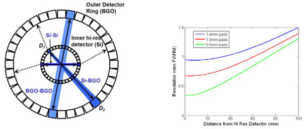
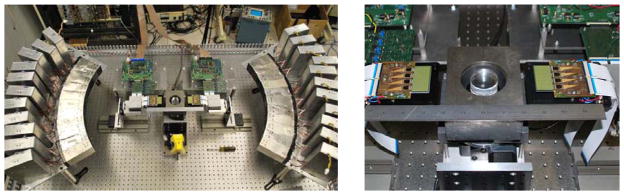
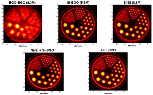
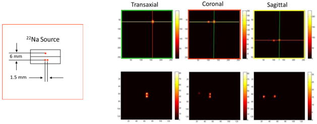
Positron emission tomography (PET) is a widely used technique in medical imaging and in studying small animal models of human disease. In the conventional approach, the 511 keV annihilation photons emitted from a patient or small animal are detected by a ring of scintillators such as LYSO read out by arrays of photodetectors. Although this has been a successful in achieving ~5mm FWHM spatial resolution in human studies and ~1mm resolution in dedicated small animal instruments, there is interest in significantly improving these figures. Silicon, although its stopping power is modest for 511 keV photons, offers a number of potential advantages over more conventional approaches. Foremost is its high spatial resolution in 3D: our past studies show that there is little diffculty in localizing 511 keV photon interactions to ~0.3mm. Since spatial resolution and reconstructed image noise trade off in a highly non-linear manner that depends on the PET instrument response, if high spatial resolution is the goal, silicon may outperform standard PET detectors even though it has lower sensitivity to 511 keV photons. To evaluate silicon in a variety of PET "magnifying glass" configurations, an instrument has been constructed that consists of an outer partial-ring of PET scintillation detectors into which various arrangements of silicon detectors can be inserted to emulate dual-ring or imaging probe geometries. Recent results have demonstrated 0.7 mm FWHM resolution using pad detectors having 16×32 arrays of 1.4mm square pads and setups have shown promising results in both small animal and PET imaging probe configurations. Although many challenges remain, silicon has potential to become the PET detector of choice when spatial resolution is the primary consideration.
Figures
References
-
- Di Domenico G, Zavattini G, Cesca N, Auricchio N, Andritschke R, Schopper F, Kanbach G. Nucl Ins Met Phys Res A. 2007;571:22–25.
-
- Park S, Rogers WL, Clinthorne NH. IEEE Trans Nucl Sci. 2007;54(5):1543–1552.
-
- Park S, Rogers WL, Clinthorne NH. Phys Med Biol. 2007;52:4653–4677. - PubMed
-
- Burdette D, Albani D, Chesi E, Clinthorne NH, Cochran E, Honscheid K, Huh SS, Kagan H, Knopp M, Lacasta C, Mikuz M, Schmallbrock P, Studen A, Weilhammer P. Nucl Ins Met Phys Res A. 2009;609:263–271.
Grants and funding
LinkOut - more resources
Full Text Sources
Other Literature Sources
Miscellaneous