

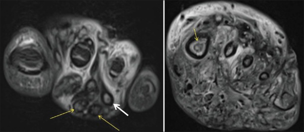
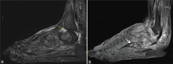
The "dot in circle" sign on MRI in maduramycosis: a characteristic finding
- PMID: 23230548
- PMCID: PMC3515922
- DOI: 10.4103/2156-7514.103056
The "dot in circle" sign on MRI in maduramycosis: a characteristic finding
Abstract
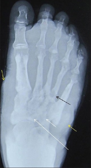
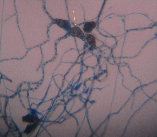
Mycetoma or Maduramycosis is a localized chronic suppurative infection characterized by exuberant granulation tissue, discharging sinuses, and bone involvement later in the course of the disease. Early clinical diagnosis before the appearance of sinuses and grains (aggregates of organism surrounded by granulation tissue, which are discharged from the draining sinuses) is difficult. Delay in diagnosis may lead to amputation of the affected part. Definitive diagnosis is through biopsy and microbiological examination. However, at times diagnosis may still be difficult. The recently described "dot in circle" sign on magnetic resonance imaging (MRI) is easy to recognize and highly specific. We present a case of mycetoma foot with characteristic MRI features.
Keywords: Dot in circle sign; MRI; maduramycosis; mycetoma.
Conflict of interest statement
Figures
References
-
- Carroll DS. Mycetoma pedis. Radiology. 1949;53:81–4. - PubMed
-
- Lewall DB, Ofole S, Bendl B. Mycetoma. Skeletal Radiol. 1985;14:257–62. - PubMed
-
- Kumar J, Kumar A, Sethy P, Gupta S. The dot-in-circle sign of mycetoma on MRI. Diagn Interv Radiol. 2007;13:193–5. - PubMed
-
- Sarris I, Berendt AR, Athanasous N, Ostlere SJ OSIRIS collaborative study group. MRI of mycetoma of the foot: Two cases demonstrating the dot-in-circle sign. Skeletal Radiol. 2003;32:179–83. - PubMed
-
- Cherian RS, Betty M, Manipadam MT, Cherian VM, Poonnoose PM, Oommen AT, et al. The “dot-in-circle” sign - A characteristic MRI finding in mycetoma foot: A report of three cases. Br J Radiol. 2009;82:662–5. - PubMed
Publication types
LinkOut - more resources
Full Text Sources
