. 2012 Dec 11:9:105.
doi: 10.1186/1742-4690-9-105.

SAMHD1 restricts HIV-1 infection in dendritic cells (DCs) by dNTP depletion, but its expression in DCs and primary CD4+ T-lymphocytes cannot be upregulated by interferons

Affiliations

SAMHD1 restricts HIV-1 infection in dendritic cells (DCs) by dNTP depletion, but its expression in DCs and primary CD4+ T-lymphocytes cannot be upregulated by interferons

Corine St Gelais et al. Retrovirology. .

Abstract

Background: SAMHD1 is an HIV-1 restriction factor in non-dividing monocytes, dendritic cells (DCs), macrophages, and resting CD4+ T-cells. Acting as a deoxynucleoside triphosphate (dNTP) triphosphohydrolase, SAMHD1 hydrolyzes dNTPs and restricts HIV-1 infection in macrophages and resting CD4+ T-cells by decreasing the intracellular dNTP pool. However, the intracellular dNTP pool in DCs and its regulation by SAMHD1 remain unclear. SAMHD1 has been reported as a type I interferon (IFN)-inducible protein, but whether type I IFNs upregulate SAMHD1 expression in primary DCs and CD4+ T-lymphocytes is unknown.

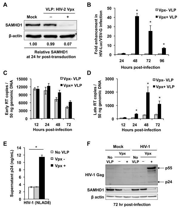
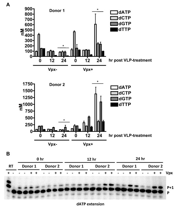
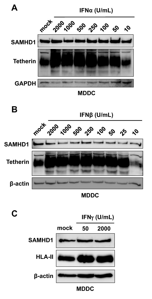
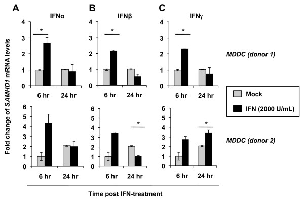
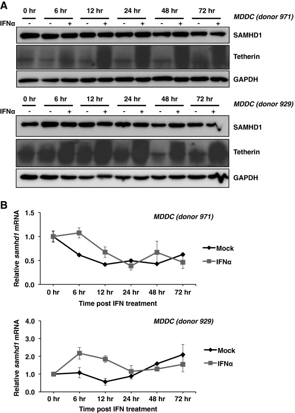
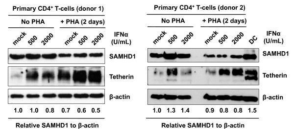
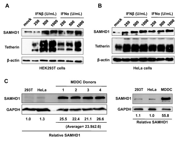
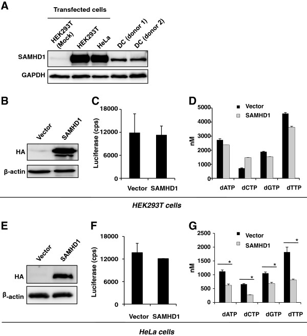
Results: Here, we report that SAMHD1 significantly blocked single-cycle and replication-competent HIV-1 infection of DCs by decreasing the intracellular dNTP pool and thereby limiting the accumulation of HIV-1 late reverse transcription products. Type I IFN treatment did not upregulate endogenous SAMHD1 expression in primary DCs or CD4+ T-lymphocytes, but did in HEK 293T and HeLa cell lines. When SAMHD1 was over-expressed in these two cell lines to achieve higher levels than that in DCs, no HIV-1 restriction was observed despite partially reducing the intracellular dNTP pool.

Conclusions: Our results suggest that SAMHD1-mediated reduction of the intracellular dNTP pool in DCs is a common mechanism of HIV-1 restriction in myeloid cells. Endogenous expression of SAMHD1 in primary DCs or CD4+ T-lymphocytes is not upregulated by type I IFNs.


Figures

Figure 1
Figure 1
Vpx-mediated SAMHD1 degradation in DCs efficiently increases HIV-1 infection and accumulation of late reverse-transcription products. Monocyte-derived DCs were transduced with SIV VLPs containing HIV-2 Vpx (Vpx +) or not (Vpx -). Mock transduction (no VLPs) was used as a negative control. (A) Whole cell lysates were subjected to immunoblotting for SAMHD1 at 24 hr post-transduction. β-actin was used as a loading control. Relative levels of SAMHD1 compared to β-actin are shown. (B) VLP-transduced DCs were infected with HIV-Luc/VSV-G at a multiplicity of infection (MOI) of 1 and the infection was detected by measuring luciferase activity in the cell lysates at the indicated times post-infection. Fold enhancement of HIV-1 infection (VLP without Vpx control set to 1) is shown. (C) HIV-1 early reverse-transcription products (early RT) in DCs transduced with VLPs. The early reverse-transcription copies were measured by qPCR at the indicated times post-HIV-1 infection (MOI of 1). (D) Increased HIV-1 late reverse-transcription products (late RT) in DCs transduced with VLPs. The late reverse-transcription copies were measured by qPCR at the indicated times post-HIV-1 infection (MOI of 1). (E and F) VLP-transduced DCs were infected with replication-competent, R5-tropic, HIV-1NLAD8 (MOI of 0.5). At 3 days post-infection, HIV-1 p24 in the supernatant was measured by ELISA. (F) Expression of SAMHD1, HIV-1 Gag (p55 and p24), and β-actin in NLAD8 HIV-1 infected DCs was detected by immunoblotting. The data shown represent one of three independent experiments. Error bars represent standard deviation of the mean of duplicate samples. (B, D, and E) The asterisks indicate a significant difference (p <0.05) compared with the controls of no VLP and/or (Vpx -).
Figure 2
Figure 2
Vpx-treatment increases the intracellular dNTP pool in DCs. Monocyte-derived DCs treated with SIVmac VLPs with and without Vpx were collected at 0, 12, and 24 hours post VLP treatment. (A) The intracellular dNTP pool of VLP-transduced DCs was measured using the single nucleotide incorporation assay. The data show results of DCs from two independent healthy donors. Error bars represent standard deviation of the mean of duplicate samples. The asterisks indicate a significant difference (p <0.001) compared with the (Vpx -) controls at 24 hr post VLP-transduction. (B). A representative HIV-1 RT-based single nucleotide primer extension gel is shown for dATP extension for both donors. Reactions (+/−) RT were used to indicate unextended primer (P) versus and extended primer (P+1). Vpx(+) VLP treated samples at 24 hr were diluted 1:3 to prevent complete primer extension and maintain results within the linear range for quantification purposes.
Figure 3
Figure 3
Type I and type II IFN treatment does not upregulate SAMHD1 expression in DCs. (A and B) Monocyte-derived DCs (MDDCs) were treated with different amounts of IFNα or IFNβ as indicated for 24 hr and whole cell lysates were subjected to immunoblotting for SAMHD1 and tetherin expression. GAPDH or β-actin was used as a loading control. (C) MDDCs were mock treated or treated with IFNγ at the indicated concentrations for 24 hours. Whole cell lysates of treated MDDCs were subjected to immunoblotting for SAMHD1 or HLA-II. β-actin was used as a loading control. The MDDC data shown represents one of two independent experiments using cells from different donors.
Figure 4
Figure 4
Type I and type II IFN treatment upregulates SAMHD1 mRNA levels in DCs at 6 hr post-treatment. Monocyte-derived DCs (MDDC) from two independent donors were either mock treated, or treated with 2000 U/mL of (A) IFNα, (B) IFNβ or (C) IFNγ as indicated for either 6 or 24 hr. Cell pellets were collected and total RNA was extracted and 200 ng of the total RNA were used for cDNA synthesis. Quantitative PCR was performed using SAMHD1 cDNA specific primers and all data was normalized to GAPDH. Data shown represent fold change in mRNA levels compared to 6 hr mock treated cells. The asterisks indicate a significant difference (p <0.05) compared with the mock controls.
Figure 5
Figure 5
Kinetics analysis of SAMHD1 protein and mRNA in DCs treated with IFNα. Monocyte-derived DCs (MDDC) from two independent donors were mock treated, indicated with “-”, or treated with 2000 U/mL IFNα, indicated with “+”, for the indicated times. (A) Whole cell lysates were subjected to immunoblotting for SAMHD1 and tetherin expression. GAPDH was used as a loading control. (B) Quantitative PCR was performed using SAMHD1 cDNA specific primers and all data was normalized to GAPDH. Data shown represents fold change in mRNA levels compared to 0 hr mock treated cells.
Figure 6
Figure 6
SAMHD1 levels in primary CD4+ T-lymphocytes are not upregulated by type I IFN treatment. Primary CD4+ T cells from two healthy donors were treated with (+) or without PHA (5 μg/mL) for 48 hr and cultured in the presence of different amounts of IFNα for 24 hr. Whole cell lysates were subjected to immunoblotting for SAMHD1 and tetherin expression. β-actin was used as a loading control and relative levels of SAMHD1 compared to β-actin are shown. The data shown represents one of three independent experiments using cells from three different donors.
Figure 7
Figure 7
Type I IFN treatment upregulates SAMHD1 expression in HEK 293T cells and HeLa cells. HEK 293T cells (A) and HeLa cells (B) were treated with different amounts of IFNα or IFNβ as indicated for 24 hr and whole cell lysates were subjected to immunoblotting for SAMHD1 and tetherin expression. β-actin is shown as a loading control. (C) Comparison of endogenous SAMHD1 expression in HEK 293T cells, HeLa cells, and monocyte-derived DCs (MDDCs) from five independent donors. Relative SAMHD1 levels compared to GAPDH are shown.
Figure 8
Figure 8
Over-expression of SAMHD1 in HEK 293T cells or HeLa cells does not inhibit HIV-1 infection, but only modestly decreases the intracellular dNTP pool. (A) HEK 293T and HeLa cells were transiently transfected with SAMHD1-expressing construct or an empty vector. SAMHD1 expression was detected by immunoblotting with SAMHD1-specific antibodies at 24 hr post-transfection. Equal amounts of total protein (20 μg) of the lysates of DCs from two donors and HEK 293T and HeLa cells were loaded. (B-D) HEK 293T cells and (E-G) HeLa cells were transiently transfected with HA-tagged SAMHD1-expressing construct or an empty vector. SAMHD1 expression in transfected HEK 293T cells (B) or HeLa cells (E) was detected by immunoblotting with anti-HA at 24 hr post-transfection. β-actin was used as a loading control. Transfected HEK 293T cells (C) or HeLa cells (F) were infected with HIV-Luc/VSV-G at an MOI of 0.5, and the infection was detected by measuring luciferase activity in the cell lysates at 48 hr post-infection and normalized to protein content (10 μg/sample). cps, counts per second. The intracellular dNTP pool of transfected HEK 293T cells (D) or HeLa cells (G) was measured at 24 hr post-transfection using the single-nucleotide incorporation assay. The asterisks indicate a significant difference (p <0.05) compared with the vector controls. The data shown represents one of two or three independent experiments. Error bars represent standard deviation of the mean of duplicate or triplicate samples.

References

    1. Coleman CM, Wu L. HIV interactions with monocytes and dendritic cells: viral latency and reservoirs. Retrovirology. 2009;6:51. doi: 10.1186/1742-4690-6-51. - DOI - PMC - PubMed
    1. Wu L, KewalRamani VN. Dendritic-cell interactions with HIV: infection and viral dissemination. Nat Rev Immunol. 2006;6(11):859–868. doi: 10.1038/nri1960. - DOI - PMC - PubMed
    1. Goujon C, Riviere L, Jarrosson-Wuilleme L, Bernaud J, Rigal D, Darlix JL, Cimarelli A. SIVSM/HIV-2 Vpx proteins promote retroviral escape from a proteasome-dependent restriction pathway present in human dendritic cells. Retrovirology. 2007;4:2. doi: 10.1186/1742-4690-4-2. - DOI - PMC - PubMed
    1. Bieniasz PD. Intrinsic immunity: a front-line defense against viral attack. Nat Immunol. 2004;5(11):1109–1115. doi: 10.1038/ni1125. - DOI - PubMed
    1. Wolf D, Goff SP. Host restriction factors blocking retroviral replication. Annu Rev Genet. 2008;42:143–163. doi: 10.1146/annurev.genet.42.110807.091704. - DOI - PMC - PubMed

Publication types

MeSH terms

Substances