

Ccl2, Cx3cr1 and Ccl2/Cx3cr1 chemokine deficiencies are not sufficient to cause age-related retinal degeneration
- PMID: 23232206
- PMCID: PMC3562441
- DOI: 10.1016/j.exer.2012.11.015
Ccl2, Cx3cr1 and Ccl2/Cx3cr1 chemokine deficiencies are not sufficient to cause age-related retinal degeneration
Abstract
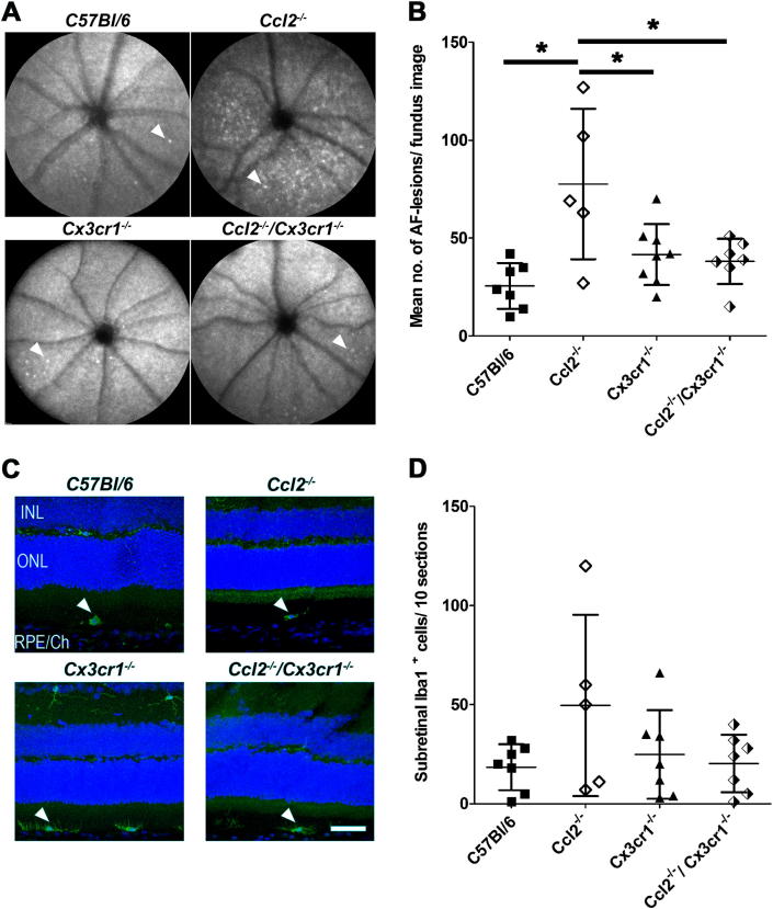
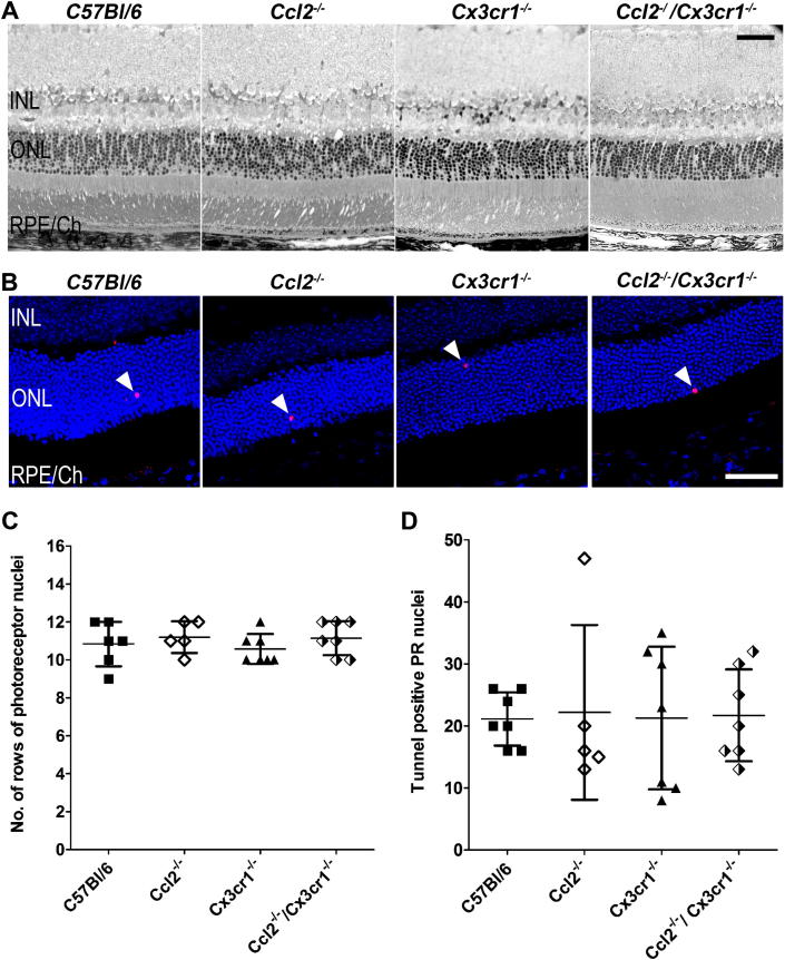
Monocytes, macrophages, dendritic cells and microglia play critical roles in the local immune response to acute and chronic tissue injury and have been implicated in the pathogenesis of age-related macular degeneration. Defects in Ccl2-Ccr2 and Cx3cl1-Cx3cr1 chemokine signalling cause enhanced accumulation of bloated subretinal microglia/macrophages in senescent mice and this phenomenon is reported to result in the acceleration of age-related retinal degeneration. The purpose of this study was to determine whether defects in CCL2-CCR2 and CX3CL1-CX3CR1 signalling pathways, alone or in combination, cause age-dependent retinal degeneration. We tested whether three chemokine knockout mouse lines, Ccl2(-/-), Cx3cr1(-/-) and Ccl2(-/-)/Cx3cr1(-/-), in comparison to age-matched C57Bl/6 control mice show differences in subretinal macrophage accumulation and loss of adjacent photoreceptor cells at 12-14 months of age. All mouse lines are derived from common parental strains and do not carry the homozygous rd8 mutation in the Crb1 gene that has been a major confounding factor in previous reports. We quantified subretinal macrophages by counting autofluorescent lesions in fundus images obtained by scanning laser ophthalmoscopy (AF-SLO) and by immunohistochemistry for Iba1 positive cells. The accumulation of subretinal macrophages was enhanced in Ccl2(-/-), but not in Cx3cr1(-/-) or Ccl2(-/-)/Cx3cr1(-/-) mice. We identified no evidence of retinal degeneration in any of these mouse lines by TUNEL staining or semithin histology. In conclusion, CCL2-CCR2 and/or CX3CL1-CX3CR1 signalling defects may differentially affect the trafficking of microglia and macrophages in the retina during ageing, but do not appear to cause age-related retinal degeneration in mice.
Copyright © 2012 Elsevier Ltd. All rights reserved.
Figures
Comment in
-
Comment on "Ccl2, Cx3cr1 and Ccl2/Cx3cr1 chemokine deficiencies are not sufficient to cause age-related retinal degeneration" by Luhmann et al. (Exp. Eye Res. 2013; 107: 80.doi: 10.1016).Exp Eye Res. 2013 Jun;111:134-5. doi: 10.1016/j.exer.2013.02.002. Epub 2013 Feb 10. Exp Eye Res. 2013. PMID: 23402809 No abstract available.
-
Reply to comment on "Ccl2, Cx3cr1 and Ccl2/Cx3cr1 chemokine deficiencies are not sufficient to cause age-related retinal degeneration" by Luhmann et al. (Exp. Eye Res. 107, February 2013, 80-87).Exp Eye Res. 2013 Jun;111:136. doi: 10.1016/j.exer.2013.02.001. Epub 2013 Feb 9. Exp Eye Res. 2013. PMID: 23402810 No abstract available.
References
-
- Ambati J., Anand A., Fernandez S., Sakurai E., Lynn B.C., Kuziel W.A., Rollins B.J., Ambati B.K. An animal model of age-related macular degeneration in senescent Ccl-2- or Ccr-2-deficient mice. Nat. Med. 2003;9:1390–1397. - PubMed
-
- Auffray C., Fogg D., Garfa M., Elain G., Join-Lambert O., Kayal S., Sarnacki S., Cumano A., Lauvau G., Geissmann F. Monitoring of blood vessels and tissues by a population of monocytes with patrolling behavior. Science. 2007;317:666–670. - PubMed
-
- Auffray C., Sieweke M.H., Geissmann F. Blood monocytes: development, heterogeneity, and relationship with dendritic cells. Annu. Rev. Immunol. 2009;27:669–692. - PubMed
-
- Cardona A.E., Pioro E.P., Sasse M.E., Kostenko V., Cardona S.M., Dijkstra I.M., Huang D., Kidd G., Dombrowski S., Dutta R., Lee J.C., Cook D.N., Jung S., Lira S.A., Littman D.R., Ransohoff R.M. Control of microglial neurotoxicity by the fractalkine receptor. Nat. Neurosci. 2006;9:917–924. - PubMed
Publication types
MeSH terms
Substances
Grants and funding
LinkOut - more resources
Full Text Sources
Other Literature Sources
Medical
Molecular Biology Databases
Research Materials
Miscellaneous
