. 2012 Dec 11;13(12):R113.
doi: 10.1186/gb-2012-13-12-r113.

Assessing telomeric DNA content in pediatric cancers using whole-genome sequencing data

Assessing telomeric DNA content in pediatric cancers using whole-genome sequencing data

Matthew Parker et al. Genome Biol. .

Abstract

Background: Telomeres are the protective arrays of tandem TTAGGG sequence and associated proteins at the termini of chromosomes. Telomeres shorten at each cell division due to the end-replication problem and are maintained above a critical threshold in malignant cancer cells to prevent cellular senescence or apoptosis. With the recent advances in massive parallel sequencing, assessing telomere content in the context of other cancer genomic aberrations becomes an attractive possibility. We present the first comprehensive analysis of telomeric DNA content change in tumors using whole-genome sequencing data from 235 pediatric cancers.

Results: To measure telomeric DNA content, we counted telomeric reads containing TTAGGGx4 or CCCTAAx4 and normalized to the average genomic coverage. Changes in telomeric DNA content in tumor genomes were clustered using a Bayesian Information Criterion to determine loss, no change, or gain. Using this approach, we found that the pattern of telomeric DNA alteration varies dramatically across the landscape of pediatric malignancies: telomere gain was found in 32% of solid tumors, 4% of brain tumors and 0% of hematopoietic malignancies. The results were validated by three independent experimental approaches and reveal significant association of telomere gain with the frequency of somatic sequence mutations and structural variations.

Conclusions: Telomere DNA content measurement using whole-genome sequencing data is a reliable approach that can generate useful insights into the landscape of the cancer genome. Measuring the change in telomeric DNA during malignant progression is likely to be a useful metric when considering telomeres in the context of the whole genome.


Figures
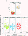
Figure 1
Figure 1
Association with matched non-tumor telomeric DNA content and age. (a) Comparison of age distribution of pediatric PCGP samples and adult TCGA samples. Because of the narrow range of age distribution in pediatric cancer in PCGP (n = 235, median = 7.5) we included 13 samples from TCGA (n = 13, median = 56) to enable evaluation of association between telomere length with age. (b) Comparison of distribution of normalized telomere count in matched normal DNA of pediatric patients with that of the adult patients. The reduction of telomere reads in adult is statistically significant (Wilcoxon signed rank P = 0.00046).
Figure 2
Figure 2
Telomere analysis using whole-genome sequencing data of 235 pediatric cancers. (a) Bayesian information criterion (BIC) guided clustering, which divided the ΔT values in this cohort into two clusters with equal variance. The boundary of these clusters is marked in dark blue. Using 0.01 as the threshold for significance, we defined the lower and upper boundary of ΔT as 'gain' or 'loss' of telomeric DNA. Samples that fall within these boundaries are deemed to have 'no change' in telomere status. (b) The number of structural variations in tumors with 'gain', 'loss' or 'no change' of telomere status. Tumors with ΔT gains have significantly higher number of structural variations compared with the other two groups (Mann-Whitney P = 1.07e-10; brain tumors P = 0.013, solid tumors P = 0.0002, hematopoietic malignancies P = NA (Not Applicable - no telomeric content gains detected); M, median). (c) The number of non-silent mutations in tumors with 'gain', 'loss' or 'no change' of telomere status. Tumors with ΔT gains have significantly higher number of sequence mutations compared with the other two groups (Mann-Whitney P = 3.723e-07; brain tumors P = 0.061; solid tumors P = 0.013, hematopoietic malignancies P = NA; M, median). (d) ΔT values from 235 pediatric cancers. The dotted lines correspond to the lower and upper boundary of ΔT as 'gain' or 'loss'. CBF, core-binding factor ALL; HYPO, hypodiploid ALL; INF, infant ALL; TALL, ETP-ALL; EPD, ependymoma; HGG, high-grade glioma; LGG, low-grade glioma; MB, medulloblastoma, ACT, adrenocortical carcinoma; NBL, neuroblastoma; OS, osteosarcoma; RB, retinoblastoma; RHB, rhabdomyosarcoma.
Figure 3
Figure 3
Age distribution versus telomere status. Age distribution in samples with different telomere status in tumor, that is, gain, loss and no change. There is no significant difference in age distribution across the three groups (ANNOVA, P = 0.368).
Figure 4
Figure 4
Validation of WGS telomeric DNA content predictions. (a) Quantitative PCR for a subset of samples, including 16 medulloblastoma and 11 neuroblastoma samples, showing changes in telomeric DNA content between normal and diagnosis log2(Absolute telomere length D/Absolute telomere length N. (b) FISH using probes for telomeric DNA confirms the 'normal' and 'abnormal' (ultra-bright spots, white arrowheads) telomere patterns in SJMB028 and SJMB004, respectively. (c) Telomere restriction fragment (TRF) analysis by southern blotting in 2 osteosarcoma samples, SJOS002 and SJOS004 predicted to have telomere gain and loss by WGS, respectively. (N = matched normal DNA, D = diagnosis tumor DNA). Inlay is the 100ng of genomic DNA on a 0.5% ethidium bromide gel which shows that the DNA quality is acceptable. (d) WGS normalized telomeric read counts for SJOS002 and SJOS004. (e) qPCR measurement of absolute telomeric DNA in SJOS002 and SJOS004 (error bars represent standard deviation of three technical repeats).

References

    1. Okuda K, Bardeguez A, Gardner JP, Rodriguez P, Ganesh V, Kimura M, Skurnick J, Awad G, Aviv A. Telomere length in the newborn. Pediatr Res. 2002;52:377–381. doi: 10.1203/00006450-200209000-00012. - DOI - PubMed
    1. d'Adda di Fagagna F, Reaper PM, Clay-Farrace L, Fiegler H, Carr P, Von Zglinicki T, Saretzki G, Carter NP, Jackson SP. A DNA damage checkpoint response in telomere-initiated senescence. Nature. 2003;426:194–198. doi: 10.1038/nature02118. - DOI - PubMed
    1. Shay JW, Bacchetti S. A survey of telomerase activity in human cancer. Eur J Cancer. 1997;33:787–791. doi: 10.1016/S0959-8049(97)00062-2. - DOI - PubMed
    1. Bryan TM, Marusic L, Bacchetti S, Namba M, Reddel RR. The telomere lengthening mechanism in telomerase-negative immortal human cells does not involve the telomerase RNA subunit. Hum Mol Genet. 1997;6:921–926. doi: 10.1093/hmg/6.6.921. - DOI - PubMed
    1. Cesare AJ, Reddel RR. Telomere uncapping and alternative lengthening of telomeres. Mech Ageing Dev. 2008;129:99–108. doi: 10.1016/j.mad.2007.11.006. - DOI - PubMed

Publication types