. 2013 Mar;56(3):654-62.
doi: 10.1007/s00125-012-2789-5. Epub 2012 Dec 12.

The role of arginase I in diabetes-induced retinal vascular dysfunction in mouse and rat models of diabetes

Affiliations

The role of arginase I in diabetes-induced retinal vascular dysfunction in mouse and rat models of diabetes

S C Elms et al. Diabetologia. 2013 Mar.

Abstract

Aims/hypothesis: A reduction in retinal blood flow occurs early in diabetes and is likely to be involved in the development of diabetic retinopathy. We hypothesise that activation of the arginase pathway could have a role in the vascular dysfunction of diabetic retinopathy.

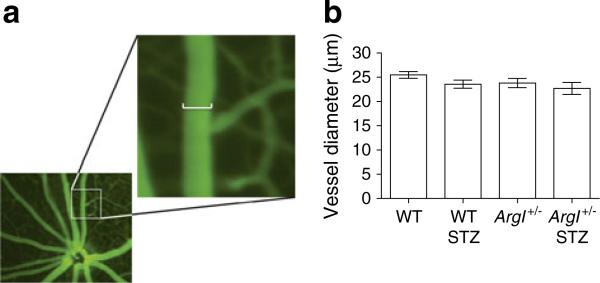
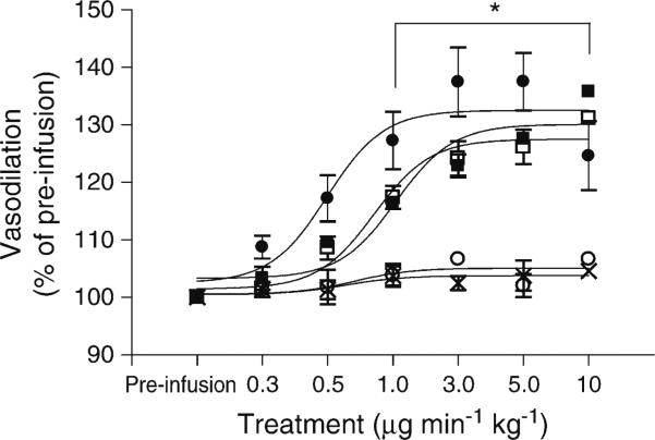
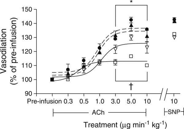
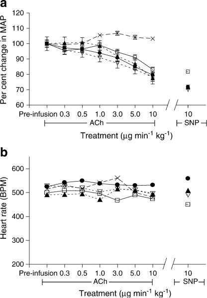
Methods: Experiments were performed using a mouse and rat model of streptozotocin (STZ)-induced diabetes for in vivo and ex vivo analysis of retinal vascular function. For in vivo studies, mice were infused with the endothelial-dependent vasodilator acetylcholine (ACh) or the endothelial-independent vasodilator sodium nitroprusside (SNP), and vasodilation was assessed using a fundus microscope. Ex vivo assays included pressurised vessel myography, western blotting and arginase activity measurements.

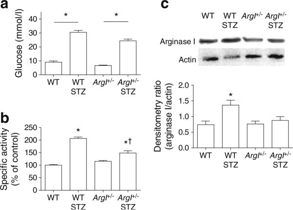
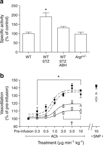
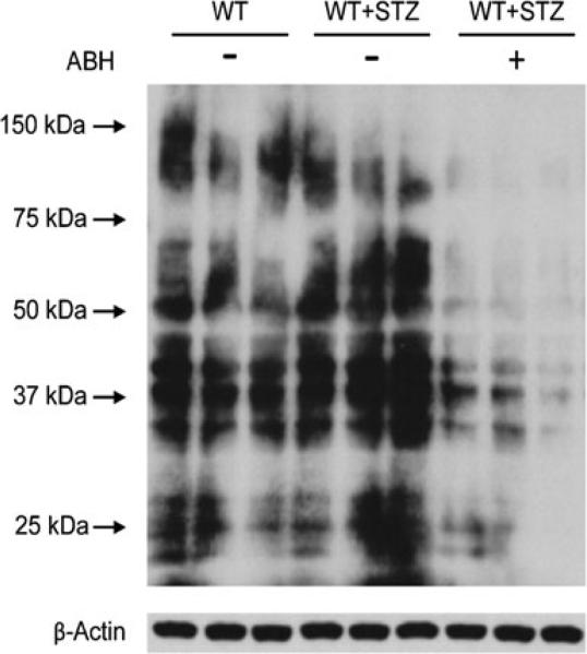
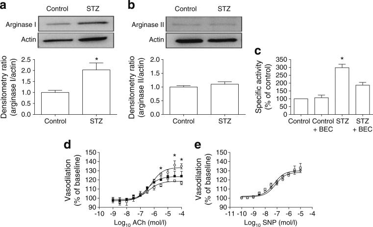
Results: ACh-induced retinal vasodilation was markedly impaired in diabetic mice (40% of control values), whereas SNP-induced dilation was not altered. The diabetes-induced vascular dysfunction was markedly blunted in mice lacking one copy of the gene encoding arginase I and in mice treated with the arginase inhibitor 2(S)-amino-6-boronohexanoic acid. Ex vivo studies performed using pressure myography and central retinal arteries isolated from rats with STZ-induced diabetes showed a similar impairment of endothelial-dependent vasodilation that was partially blunted by pretreatment of the isolated vessels with another arginase inhibitor, (S)-2-boronoethyl-L-cysteine. The diabetes-induced vascular alterations were associated with significant increases in both arginase I protein levels and total arginase activity.

Conclusions/interpretation: These results indicate that, in the mouse and rat model, diabetes-induced increases in arginase I were involved in the diabetes-induced impairment of retinal blood flow by a mechanism involving vascular endothelial cell dysfunction.


Figures

Fig. 1
Fig. 1
Blood glucose, arginase activity and arginase I protein levels assessed as described in the Methods section. (a) WT STZ and ArgI+/– STZ mice had significantly higher blood glucose levels than the non-diabetic controls (*p<0.05 vs all groups, p<0.05 vs WT STZ, oneway ANOVA; WT, n=9; WT STZ, n=6; ArgI+/–, n=10; ArgI+/– STZ, n=6). (b) WT STZ and ArgI+/– STZ retinas showed significantly greater arginase activity than those from non-diabetic controls (*p<0.05, oneway ANOVA; all groups, n=4). (c) WT STZ retinas showed higher levels of ArgI protein compared with WT and ArgI+/– retinas. Actin protein was used for loading control. (*p<0.05, one-way ANOVA; WT, n=9; WT STZ, n=8; ArgI+/–, n=11; ArgI+/– STZ, n=4)
Fig. 2
Fig. 2
Diameter of the secondary retinal arterioles measured using the Micron III fundoscope as described in the Methods section. (a) Measurements were taken from fully magnified images in which one pixel=2.25 μm. The scale bar in the enlargement represents 12 pixels (27 μm). (b) Vessel diameter measurements taken pre-infusion (at baseline) showed no differences between the groups (one-way ANOVA; WT, n=31; WT STZ, n=12; ArgI+/–, n=20; ArgI+/– STZ, n=16)
Fig. 3
Fig. 3
Fundus imaging of the retinal vasculature in non-diabetic WT mice showed that the retinal vessels dilated equally to SNP and ACh at maximal dilation. Pretreatment with the muscarinic receptor blocker atropine completely blocked dilation to ACh at all doses, but had no effect on SNP-mediated dilation. The vehicle had no effect on retinal vasodilation. (*p<0.05 WT ACh + atropine compared with all other groups, two-way ANOVA; all groups, n=5). Black circle, WT ACh; black square, WT SNP; white circle, WT ACh + atropine; white square, WT SNP + atropine; cross, vehicle
Fig. 4
Fig. 4
Fundus imaging of the retinal vasculature of STZ-diabetic and age-matched control WT and ArgI+/– mice, showing that vasodilation to ACh was markedly reduced in the vessels of WT STZ mice compared with the other three groups. ACh-induced dilation was significantly enhanced in the ArgI+/– STZ mice compared with the WT STZ mice. All four groups responded similarly to SNP (*p<0.05 WT and ArgI+/– compared with WT STZ; p<0.05 ArgI+/– STZ compared with WT STZ, two-way ANOVA; WT, WT STZ and ArgI+/–, n=4; ArgI+/– STZ, n=6). Black circle, WT; white square, WT STZ; black triangle, ArgI+/–; white triangle, ArgI+/– STZ
Fig. 5
Fig. 5
Blood pressure and HR were not altered by diabetes or heterozygous deletion of arginase I. (a) ACh and SNP elicited equal depressant effects on blood pressure in all groups. (b) ACh and SNP had no effect on HR over the entire course of infusion, and there were no differences between the groups. The vehicle had no effect on either MAP or HR (all groups, n=4). Black circle, WT; white square, WT STZ; black triangle, ArgI+/–; white triangle, ArgI+/– STZ; cross, vehicle
Fig. 6
Fig. 6
Treatment with the specific arginase inhibitor ABH reduced arginase activity and improved vasodilation in the WT STZ retinas. (a) Arginase activity was significantly reduced in the ABH-treated WT STZ retinas compared with the untreated WT STZ retinas (*p<0.05 WT STZ compared with all other groups, one-way ANOVA; WT, n=4; WT STZ, n=5; WT STZ ABH, n=6; ArgI+/–, n=5). (b) Dilation to ACh was significantly improved in the ABH-treated mice compared with the WT STZ mice (*p<0.05 WT and ArgI+/– compared with WT STZ; p<0.05 WT STZ ABH compared with WT STZ, two-way ANOVA; WT, n=6; WT STZ, n=4; WT STZ ABH, n=10; ArgI+/–, n=4). Black circle, WT; white square, WT STZ; white triangle, WT STZ ABH; black triangle, ArgI+/–
Fig. 7
Fig. 7
Representative western blot for 3-nitrotyrosine showed greater 3-nitrotyrosine immunoreactivity in the retinas from STZ mice than those from control mice. The arginase inhibitor ABH blocked the formation of 3-nitrotyrosine in the STZ mice. Each experimental group consists of three lanes containing one retina per lane. β-Actin was used as the loading control
Fig. 8
Fig. 8
Diabetes-induced increases in arginase I protein levels and arginase activity in the STZ rat retina were associated with impaired vasodilation of the CRA as shown by ex vivo pressure myography. (a, b) Western blots and densitometry show STZ-induced upregulation of ArgI (a) but not ArgII (b) compared with actin loading controls. (c) Arginase activity was significantly increased in the STZ CRAs compared with the controls (*p<0.05). This increase was significantly blunted by ex vivo treatment of the isolated vessels with the arginase inhibitor BEC (*p<0.05, one-way ANOVA; all groups, n=4). (d) Vasodilation in the CRAs from STZ rats was reduced compared with controls and with STZ vessels pretreated with BEC (*p<0.05, twoway ANOVA; all groups, n=3). (e) Dilation of CRAs to SNP was no different in the STZ diabetic rats and the age-matched controls. White circle, controls; white square, STZ; black square, STZ BEC

References

    1. Curtis TM, Gardiner TA, Stitt AW. Microvascular lesions of diabetic retinopathy: clues towards understanding pathogenesis? Eye (Lond) 2009;23:1496–1508. - PubMed
    1. Clermont AC, Bursell SE. Retinal blood flow in diabetes. Microcirculation. 2007;14:49–61. - PubMed
    1. Nagaoka T, Sato E, Takahashi A, Yokota H, Sogawa K, Yoshida A. Impaired retinal circulation in patients with type 2 diabetes mellitus: retinal laser Doppler velocimetry study. Invest Ophthalmol Vis Sci. 2010;51:6729–6734. - PubMed
    1. De Caterina R, Libby P, Peng HB, et al. Nitric oxide decreases cytokine-induced endothelial activation. Nitric oxide selectively reduces endothelial expression of adhesion molecules and proinflammatory cytokines. J Clin Invest. 1995;96:60–68. - PMC - PubMed
    1. Furchgott RF, Zawadzki JV. The obligatory role of endothelial cells in the relaxation of arterial smooth muscle by acetylcholine. Nature. 1980;288:373–376. - PubMed

Publication types

LinkOut - more resources