. 2013 Feb;19(1):75-81.
doi: 10.1007/s13365-012-0142-x. Epub 2012 Dec 12.

Varicella zoster virus infection of highly pure terminally differentiated human neurons

Affiliations

Varicella zoster virus infection of highly pure terminally differentiated human neurons

Xiaoli Yu et al. J Neurovirol. 2013 Feb.

Abstract

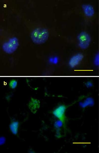
In vitro analyses of varicella zoster virus (VZV) reactivation from latency in human ganglia have been hampered by the inability to isolate virus by explantation or cocultivation techniques. Furthermore, attempts to study interaction of VZV with neurons in experimentally infected ganglion cells in vitro have been impaired by the presence of nonneuronal cells, which become productively infected and destroy the cultures. We have developed an in vitro model of VZV infection in which highly pure (>95 %) terminally differentiated human neurons derived from pluripotent stem cells were infected with VZV. At 2 weeks post-infection, infected neurons appeared healthy compared to VZV-infected human fetal lung fibroblasts (HFLs), which developed a cytopathic effect (CPE) within 1 week. Tissue culture medium from VZV-infected neurons did not produce a CPE in uninfected HFLs and did not contain PCR-amplifiable VZV DNA, but cocultivation of infected neurons with uninfected HFLs did produce a CPE. The nonproductively infected neurons contained multiple regions of the VZV genome, as well as transcripts and proteins corresponding to VZV immediate-early, early, and late genes. No markers of the apoptotic caspase cascade were detected in healthy-appearing VZV-infected neurons. VZV infection of highly pure terminally differentiated human neurons provides a unique in vitro system to study the VZV-neuronal relationship and the potential to investigate mechanisms of VZV reactivation.


Figures

Fig. 1
Fig. 1
VZV infection of highly pure human neurons did not produce a cytopathic effect. Terminally differentiated neurons were maintained in tissue culture for up to 21 days. Phase-contrast microscopy showed healthy-appearing neurons on day 0 (a) and day 14 in culture (b) as well as 14 days after VZV infection (c). Dual immunofluorescence staining (d–f) with anti-βIII-tubulin antibody and anti-GFAP antibody revealed positive staining for the neuronal marker tubulin (red), but not for GFAP (green). Nuclei stained blue with DAPI
Fig. 2
Fig. 2
VZV DNA was present in non-productively infected human neurons. Two weeks after VZV infection, DNA was extracted from neurons and quantitated by real-time PCR. Multiple regions of the VZV genome were detected (black and gray bars are VZV and GAPdH DNA, respectively)
Fig. 3
Fig. 3
Multiple VZV transcripts were present in non-productively infected neurons. Two weeks after infection of neurons, total RNA was extracted, treated with DNAse, and cDNA was synthesized. Transcripts corresponding to VZV immediate early (IE), early (E) and late (L) genes were quantified by qPCR as compared to RNA obtained from VZV-infected fibroblasts at the height of a cytopathic effect (black and gray bars are neurons and HLF, respectively)
Fig. 4
Fig. 4
VZV immediate-early proteins were present in non-productively infected human neurons. Two weeks after VZV infection, neurons attached to laminin-coated coverslips were fixed and immunostained with anti-tubuiln antibody and anti-VZV IE 62 antibody. Highly pure, βIII-tubulin-positive human neurons (red, a) contain VZV IE62 in the nucleus (green). (b and c) show dual immunostained neurons: VZV IE62 protein was seen in the nucleus (green) and VZV IE 63 in the cytoplasm (red). Scale bar: 20 μm
Fig. 5
Fig. 5
VZV early proteins were present in non-productively infected human neurons. Two weeks after VZV infection, neurons attached to laminin-coated coverslips were fixed and immunostained with anti-VZV ORF 29 antibody and anti-VZV thymidine kinase (TK) antibody. (Fig. 5a) shows dual immunostaining of DAPI and early VZV ORF29 protein in the nucleus (green). (Fig. 5b) shows dual immunostaining of DAPI and anti-VZV TK antibody. VZV TK was seen in the cytoplasm of infected neurons. Scale bars: 20 μm
Fig. 6
Fig. 6
VZV late proteins gE and gH were detected in non-productively infected human neurons. Two weeks after VZV infection, neurons attached to laminin-coated coverslips were fixed and immunostained with anti-VZV gE (a) or anti-VZV gH (b). Dual immunostaining revealed late VZV gE in the cytoplasm (green, a) and VZV gH in the cytoplasmic membrane (green, b). Scale bars: 20 μm
Fig. 7
Fig. 7
Caspase-3 and caspase-9 proteins were not detected in VZV-infected neurons. Two weeks after VZV infection, neurons attached to laminin-coated coverslips were fixed and dual-immunostained with anti-caspase 3 (red) and anti-VZV 62 (green) or with anti-caspase 9 (red) and anti-VZV 62 (green) antibodies. Figs. 7a and c show VZV 62 protein (green) but not caspase-3 (red) or caspase-9 (red) in non-productively infected neurons; Figs. 7b and d show extensive caspase-3 (red) and caspase-9 (red) and VZV IE 62 (green) staining in VZV-infected fibroblasts

References

    1. Baiker A, Fabel K, Cozzio A, Zerboni L, Fabel K, Sommer M, Uchida N, He D, Weissman I, Arvin AM. Varicella-zoster virus infection of human neural cells in vivo. Proc Natl Acad Sci USA. 2004;101:10792–10797. doi: 10.1073/pnas.0404016101. - PMC - PubMed
    1. Christensen J, Steain M, Slobedman B, Abendroth A. Differentiated neuroblastoma cells provide a highly efficient model for studies of productive varicella-zoster virus infection of neuronal cells. J Virol. 2011;85:8436–8442. - PMC - PubMed
    1. Dukhovny A, Sloutskin A, Markus A, Yee MB, Kinchington PR, Goldstein RS. Varicella-zoster virus infects human embryonic stem cell-derived neurons and neurospheres but not pluripotent embryonic stem cells or early progenitors. J Virol. 2012;86:3211–3218. - PMC - PubMed
    1. Hood C, Cunningham AL, Slobedman B, Boadle RA, Abendroth A. Varicella-zoster virus-infected human sensory neurons are resistant to apoptosis, yet human foreskin fibroblasts are susceptible: evidence for a cell-type-specific apoptotic response. J Virol. 2003;77:12852–12864. doi: 10.1128/JVI.77.23.12852-12864. - PMC - PubMed
    1. Gilden DH, Cohrs RJ, Mahalingam R. Clinical and molecular pathogenesis of varicella virus infection. Viral Immunol. 2003;16:243–258. - PubMed

Publication types