

doi: 10.1002/anie.201207063.
Epub 2012 Dec 11.
DNA aptamer-mediated cell targeting
Affiliations
- PMID: 23233389
- PMCID: PMC3793636
- DOI: 10.1002/anie.201207063
Item in Clipboard
DNA aptamer-mediated cell targeting
Angew Chem Int Ed Engl.
.
Abstract
One important issue using cells as therapeutics is targeted delivery. Engineering cell surfaces to improve delivery efficiency is thus of great interest. Here we report a simple, efficient and effective way to modify the cell surface with target-specific ligands, i.e., DNA aptamers, while minimizing the effects on the modified cells. We demonstrated that after incubating with lipo-aptamer probes (shown in expansion), immune cells (red) recognize cancer cells (blue) in the cell mixture, and kill cancer cells.
Figures
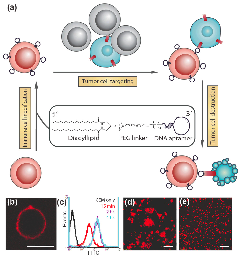
Modification of cell membranes with aptamers. (a) Schematic representation of targeting cancer cells (blue) with aptamer-modified immune cells (red). After incubating with lipo-aptamer probes (shown in expansion), immune cells recognize cancer cells in the cell mixture, and kill cancer cells. (b) Confocal microscope image of lipo-Lib-TMR-treated CEM cells. Red fluorescent probes were found only on the cell surface. Scale bar: 10 µm. (c) CEM cells were treated with lipo-Lib-FITC for different time intervals in cell culture medium. The maximum insertion was reached in 2 hours. (d) Ramos cells spontaneously aggregate after treatment with lipo-TD05-TMR. Scale bar: 100 µm. (e) Control experiments showed no assembly when Ramos cells were treated with lipo-lib-TMR. Scale bar: 100 µm.
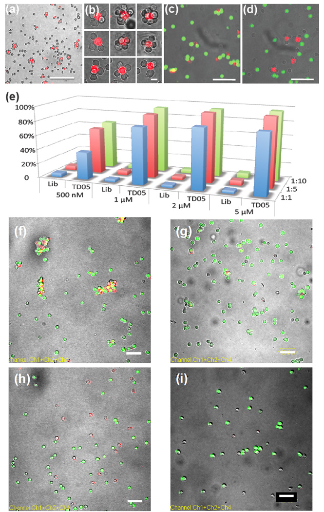
Aptamer-directed assembly and disassembly of cell aggregates and quantitative analysis of aggregation. (a) 1:10 mixture of lipo-sgc8-TMR-modified Ramos and CEM cells. Scale bar: 100 µm. (b) Discrete cell clusters at higher magnification. Scale bar: 20 µm. (c) Confocal micrograph of selective cell assembly. Lipo-TD05-TMR-modified K562 cells (red fluorescence) were incubated with CellTracker Green-labeled Ramos cells (green fluorescence) and unlabeled CEM cells. No mismatched cell assembly (K562 and CEM cells or Ramos and CEM cells) was observed. Scale bar: 50 µm. (d) Confocal micrograph of selective cell assembly of lipo-Sgc8-TMR-modified K562 cells (red fluorescence) and unlabeled CEM cells. (e) Aggregation percentage of CEM cells in different sample sets. CEM-to-Ramos cell ratios are represented by blue (1:1), red (1:5) and green (1:10) bars. (f) 1:5 mixture of lipo-TD05-TMR-modified CEM (red) cells and Ramos (green) cells after 20 min incubation. CEM and Ramos cells formed aggregates. Scale bar: 100 µm. (g) Mixture of Lipo-TD05-TMR-modified CEM (red) cells and Ramos (green) cells from (f) treated with DNase I. Scale bar: 100 µm. (h) 1:5 mixture of lipo-Lib-TMR-modified CEM (red) cells and Ramos (green) cells after 25 min incubation. CEM and Ramos cells remained apart. Scale bar: 100 µm. (i) Mixture of Lipo-Lib-TMR-modified CEM (red) cells and Ramos (green) cells from (h) treated with DNase I. Scale bar: 100 µm.
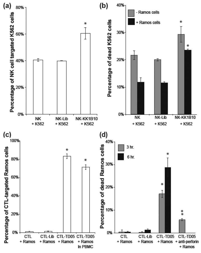
Aptamer-assisted immune effector cell targeting and killing of leukemia cells (a) NK-K562 cell binding assay. NK cells recognize K562 cells spontaneously; however, aptamer modification improved the targeting efficiency by 50%. (b) NK-K562 cell killing assay. Aptamer-modified NK cells killed K562 cells more efficiently, especially in the presence of background cells (Ramos). (c) CTL-Ramos cell binding assay. Without presenting the specific MHC1:peptide on Ramos cell surfaces, CTL cannot recognize Ramos cells. On the other hand, aptamer-modified CTL recognized 80% Ramos cells. (d) CTL-Ramos cell killing assay. Assisted by membrane-anchored aptamers, CTL targeted and killed Ramos cells via cell-mediated immunity. CTL-mediated cytotoxicity was confirmed by blocking the perforin/granzyme pathway with antibody. Values are means with SD (n=3). The single asterisk indicates a significant difference between aptamer-modified and unmodified or Lib-modified groups determined by the one-tailed t-test at P<0.01. The double asterisks indicate a significant difference between aptamer-modified and anti-Perforin treated groups determined by the one-tailed t-test at P<0.01.
References
-
- van der Bruggen P, Traversari C, Chomez P, Lurquin C, De Plaen E, Van den Eynde B, Knuth A, Boon T. Journal of Immunology. 2007;178:2617–2621. - PubMed
- Kawakami Y, Eliyahu S, Delgado CH, Robbins PF, Sakaguchi K, Appella E, Yannelli JR, Adema GJ, Miki T, Rosenberg SA. Proceedings of the National Academy of Sciences of the United States of America. 1994;91:6458–6462. - PMC - PubMed
- Albertsson PA, Basse PH, Hokland M, Goldfarb RH, Nagelkerke JF, Nannmark U, Kuppen PJK. Trends in Immunology. 2003;24:603–609. - PubMed
-
- Conrad C, Gupta R, Mohan H, Niess H, Bruns CJ, Kopp R, von Luettichau I, Guba M, Heeschen C, Jauch K-W, Huss R, Nelson PJ. Current Gene Therapy. 2007;7:249–260. - PubMed
- Reiser J, Zhang XY, Hemenway CS, Mondal D, Pradhan L, La Russa VF. Expert Opinion on Biological Therapy. 2005;5:1571–1584. - PMC - PubMed
- Elzaouk L, Moelling K, Pavlovic J. Experimental Dermatology. 2006;15:865–874. - PubMed
-
- Foxall C, Watson SR, Dowbenko D, Fennie C, Lasky LA, Kiso M, Hasegawa A, Asa D, Brandley BK. Journal of Cell Biology. 1992;117:895–902. - PMC - PubMed
- Sarkar D, Spencer JA, Phillips JA, Zhao WA, Schafer S, Spelke DP, Mortensen LJ, Ruiz JP, Vemula PK, Sridharan R, Kumar S, Karnik R, Lin CP, Karp JM. Blood. 2011;118:E184–E191. - PMC - PubMed
Publication types
MeSH terms
Substances
Grants and funding
LinkOut - more resources
Full Text Sources
Other Literature Sources
