. 2013 Mar;12(3):626-37.
doi: 10.1074/mcp.M112.021931. Epub 2012 Dec 11.

A proteomics and transcriptomics approach to identify leukemic stem cell (LSC) markers

Affiliations

A proteomics and transcriptomics approach to identify leukemic stem cell (LSC) markers

Francesco Bonardi et al. Mol Cell Proteomics. 2013 Mar.

Abstract

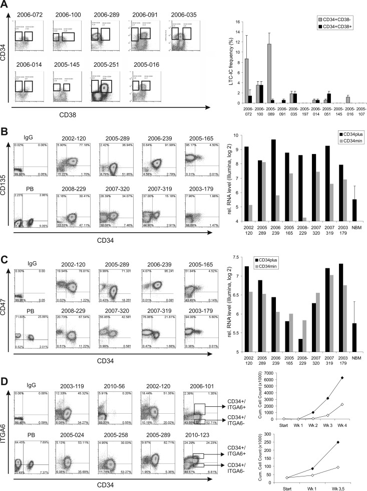
Interactions between hematopoietic stem cells and their niche are mediated by proteins within the plasma membrane (PM) and changes in these interactions might alter hematopoietic stem cell fate and ultimately result in acute myeloid leukemia (AML). Here, using nano-LC/MS/MS, we set out to analyze the PM profile of two leukemia patient samples. We identified 867 and 610 unique CD34(+) PM (-associated) proteins in these AML samples respectively, including previously described proteins such as CD47, CD44, CD135, CD96, and ITGA5, but also novel ones like CD82, CD97, CD99, PTH2R, ESAM, MET, and ITGA6. Further validation by flow cytometry and functional studies indicated that long-term self-renewing leukemic stem cells reside within the CD34(+)/ITGA6(+) fraction, at least in a subset of AML cases. Furthermore, we combined proteomics with transcriptomics approaches using a large panel of AML CD34(+) (n = 60) and normal bone marrow CD34(+) (n = 40) samples. Thus, we identified eight subgroups of AML patients based on their specific PM expression profile. GSEA analysis revealed that these eight subgroups are enriched for specific cellular processes.


Conflict of interest statement

Conflict of interest: The authors declare no conflict of interest.

Figures

Fig. 1.
Fig. 1.
Identification of the plasma membrane proteome of leukemic stem cell-enriched fractions of primary leukemia patient samples. A, Schematic workflow of the proteome strategy adopted for the AML samples analysis. B, Venn diagram showing the number of proteins identified for the two AML samples. Total refers to the total amount of identified proteins. PM+ refers to a subgroup of selected proteins included in the following GO terms: plasma membrane, extracellular region, cell projection, extrinsic to membrane, extracellular matrix. PM indicated the number of proteins annotated as plasma membrane only. C, Pie chart indicating the composition of the samples after the purification procedure indicated in panel A. D, Venn diagram showing the number of total proteins, PM+ proteins, and PM proteins identified in the CD34+ fractions of the two AML samples. E, Gene ontology annotation for biological processes using the combined list of all identified AML CD34+ PM proteins. F, Subset of PM proteins identified in AML CD34+ fractions.
Fig. 2.
Fig. 2.
Identification of leukemic stem cell markers using a transcriptomics approach. A, Scheme representing the transcriptome strategy adopted to analyze the AML and NBM samples. B, Heat map showing how the supervised clustering of the 238 differentially expressed genes obtained with the procedures indicated in panel A. C, Venn diagram showing the overlap between the plasma membrane proteins identified with the proteome approach and those identified with the transcriptome approach. The 59 overlapping proteins are further GO-annotated for molecular function.
Fig. 3.
Fig. 3.
Verification and functional characterization of a number of putative leukemic stem cell markers. A, Expression of CD34 and CD38 among 10 primary AML patients (left) and relative stem cell frequencies as determined by long-term culture-initiating cell assays (right). B, Validation of the expression of CD135, C, CD47, and (D) ITGA6, by FACS analysis (left panels) by FACS analysis. In (B) and (C), comparisons with mRNA levels obtained with Illumina BeadArray are also shown (right graphs). In two cases, CD34+/ITGA6+ and CD34+/ITGA6 populations were sorted and long-term expansion was analyzed in MS5 cocultures (D, right panels).
Fig. 4.
Fig. 4.
Evaluating heterogeneity in plasma membrane markers in AML. A, By identifying the best discriminating uncorrelated markers using an information gain approach (see “Results” section and Materials and Methods sections for details) we were able to identify eight plasma membrane markers that were almost completely uncorrelated. Supervised cluster analysis of expression of these eight markers in AML CD34+ and NBM CD34+ samples is shown. B, An overview of the eight uncorrelated markers including information gain is shown. Gene set enrichment analysis (GSEA) of the eight plasma membrane markers indicates that the identified subgroups associate with specific gene signatures. NS denotes Not Significant.

References

    1. Estey E., Döhner H. (2006) Acute myeloid leukaemia. Lancet 368, 1894–1907 - PubMed
    1. Löwenberg B., Downing J. R., Burnett A. (1999) Acute myeloid leukemia. N. Engl. J. Med. 341, 1051–1062 - PubMed
    1. Bonnet D., Dick J. E. (1997) Human acute myeloid leukemia is organized as a hierarchy that originates from a primitive hematopoietic cell. Nat. Med. 3, 730–737 - PubMed
    1. Dalerba P., Cho R. W., Clarke M. F. (2007) Cancer stem cells: models and concepts. Annu. Rev. Med. 58, 267–284 - PubMed
    1. Doulatov S., Notta F., Laurenti E., Dick J. E. (2012) Hematopoiesis: a human perspective. Cell Stem Cell 10, 120–136 - PubMed

Publication types

MeSH terms