. 2013 Mar;12(3):797-806.
doi: 10.1074/mcp.O112.024059. Epub 2012 Dec 11.

A high-throughput assay for phosphoprotein-specific phosphatase activity in cellular extracts

Affiliations

A high-throughput assay for phosphoprotein-specific phosphatase activity in cellular extracts

Anjun K Bose et al. Mol Cell Proteomics. 2013 Mar.

Abstract

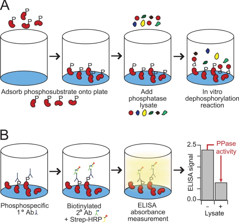
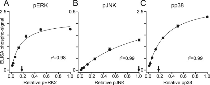
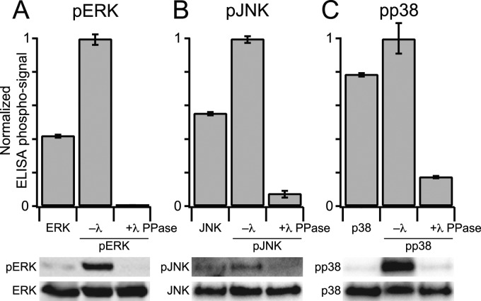
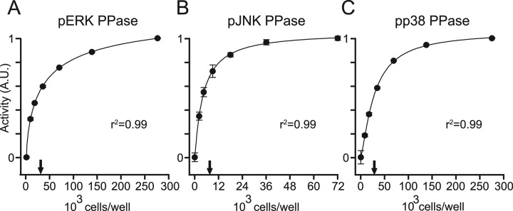
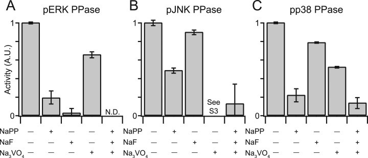
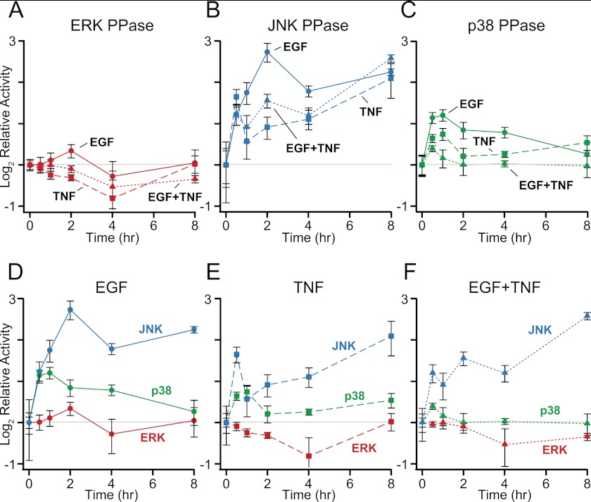
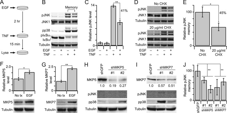
Protein phosphatases undo the post-translational modifications of kinase-signaling networks, but phosphatase activation in cells is difficult to measure and interpret. Here, we report the design of a quantitative and high-throughput assay platform for monitoring cellular phosphatase activity toward specific phosphoprotein targets. Protein substrates of interest are purified recombinantly, phosphorylated in vitro using the upstream kinase, and adsorbed to 96-well plates. Total phosphatase extracts from cells are then added to trigger a solid-phase dephosphorylation reaction. After stopping the reaction, phosphoprotein levels are quantified by ELISA with a phospho-specific antibody, and the loss of phospho-specific immunoreactivity is used as the readout of phosphatase activity. We illustrate the generality of the method by developing specific phosphatase-activity assays for the three canonical mitogen-activated protein phospho-kinases: ERK, JNK, and p38. The assays capture changes in activity with a dynamic range of 25-100-fold and are sensitive to a limit of detection below 25,000 cells. When applied to cytokine-induced signaling, the assays revealed complex and dynamic regulation of phosphatases suggesting cross-communication and a means for cellular memory. Our assay platform should be beneficial for phosphoproteomic surveys and computational-systems models of signaling, where phosphatases are known to be important but their activities are rarely measured.


Figures

Fig. 1.
Fig. 1.
A high-throughput phosphatase assay that monitors endogenous activity toward specific phosphoprotein substrates. A, recombinant phosphorylated substrate is adsorbed onto a polystyrene microtiter plate, the immobilized phosphosubstrate are treated with PPase lysate, and the dephosphorylation reaction is halted with the addition of a phosphatase inhibitor mixture. B, the remaining phosphosubstrate is labeled with anti-phosphoprotein primary antibody, biotinylated secondary antibody, and streptavidin-HRP for colorimetric detection. Phosphatase activity is determined by the drop in phosphoprotein signal in lysate-treated samples relative to negative controls.
Fig. 2.
Fig. 2.
ELISA-based quantification of recombinant pMAPKs adsorbed to 96-well plates. A–C, Twofold dilution series (relative level 1.0 = ∼400 ng) of pERK (A), pJNK (B), and pp38 (C) were adsorbed onto polystyrene microtiter plates and detected by ELISA using phospho-specific antibodies. The detection saturates at ∼75 ng for pERK (A), ∼400 ng for pJNK (B), and ∼75 ng for pp38 (C). Arrows indicate the quantity of pMAPKs used in the PPase assay. Data are shown as the mean ± S.E. of three independent assay replicates.
Fig. 3.
Fig. 3.
The ELISA measurement of pMAPK levels is phospho-specific. A–C (upper panels), immobilized MAPKs or pMAPKs were incubated in the presence (+) or absence (–) of lambda (λ) PPase and the resulting phosphorylation levels of pERK (A), pJNK (B), or pp38 (C) were measured by ELISA. A-C (lower panels), matched λ PPase-treated samples were analyzed by immunoblotting for pERK (A), pJNK (B), or pp38 (C) with Flag or total MAPK used as a loading control. Data are shown as the mean ± S.E. of three independent assay replicates.
Fig. 4.
Fig. 4.
Early reaction kinetics of the MAPK PPase activity assays are linear with time. A–C, immobilized pERK (A), pJNK (B), or pp38 (C) were treated with saturating concentrations of PPase lysate (see Fig. 5) or lysis buffer for the indicated times. Arrows indicate the incubation times used in the optimized assays. Gray markers indicate time points where the kinetics have saturated. Data are shown as the mean ± S.E. of three independent assay replicates.
Fig. 5.
Fig. 5.
MAPK PPase activity assays are dose-dependent with respect to the concentration of cell lysate, and measure activity with fewer than 25,000 cells per well. A–C, the PPase assays were performed with 2-fold serial dilutions of concentrated PPase lysate (∼275,000 to ∼9,000 cells/well for pERK (A) and pp38 (C), and ∼70,000 to ∼2,000 cells/well for pJNK (B)). Measured activities were regressed against cell lysate using a four-parameter logistic curve. Arrows indicate the concentration of lysate used in the optimized assays. Data are shown as the mean ± S.E. of three independent assay replicates.
Fig. 6.
Fig. 6.
MAPK PPase activity assays specifically report PPase enzymatic activity in cell extracts. A–C, PPase lysates were treated with 10 mm NaPP, 30 mm NaF, 200 μm Na3VO4, or a combination of all three PPase inhibitors, and PPase activity was measured toward pERK (A), pJNK (B), or pp38 (C). See supplemental Fig. S3 for an explanation of the JNK PPase assay results for Na3VO4-treated lysates. Data are shown as the mean ± S.E. of three independent assay replicates.
Fig. 7.
Fig. 7.
Cytokine stimulation evokes quantitatively and temporally distinct MAPK PPase activation profiles. HT-29 cells were seeded at 50,000 cells/cm2, sensitized with 200 U/ml IFNγ, and stimulated with 100 ng/ml EGF, 100 ng/ml TNF, or both for the indicated time points. A–C, cells were lysed and measured for PPase activity against pERK (A), pJNK (B), and pp38 (C) as described in the experimental procedures. D–F, the MAPK PPase activity measurements are replotted with respect to EGF (D), TNF (E), and EGF+TNF (F). Data are shown as the mean ± S.E. of four biological replicates.
Fig. 8.
Fig. 8.
EGF up-regulates MKP5 and MKP7 to prime cells against future JNK-activating stimuli. A, HT-29 cells were seeded at 50,000 cells/cm2, sensitized with 200 U/ml IFN-γ, and stimulated with 100 ng/ml EGF for 2 h to upregulate JNK PPase activity, followed by 100 ng/ml TNF for 15 min. B, cells with or without 100 ng/ml EGF prestimulation or 100 ng/ml TNF stimulation were analyzed for pJNK, pp38, or total IκBα levels with JNK1 and tubulin used as loading controls. pIκBα appears as an upshifted band on the total IκBα immunoblot. C, replicated densitometry of the results shown in (B). D, inhibition of protein synthesis inhibits EGF-induced pJNK memory. Cells were pretreated with 20 μg/ml cycloheximide (CHX) before EGF-TNF stimulation as described in (A). E, replicated densitometry of the results shown in (D). F and G, EGF up-regulates MKP5 and MKP7. Cells were stimulated with 100 ng/ml EGF for 2 h and analyzed for MKP5 or MKP7 by immunoblotting with tubulin used as a loading control. Replicated densitometry is shown in the upper panels. H and I, shRNA-mediated knockdown of MKP5 or MKP7. Cells were transduced with lentiviruses, selected, and analyzed for MKP5 or MKP7, pJNK, or pp38 with tubulin used as a loading control. Relative densitometry for the extent of knockdown is shown beneath the MKP5 and MKP7 immunoblots. J, Knockdown of MKP5 or MKP7 reduces EGF-induced pJNK memory. shMKP5 or shMKP7 cells were stimulated with EGF-TNF as described in (A) and analyzed for pJNK by immunoblotting. Replicated densitometry is shown compared with shGFP control cells. Data are shown as the mean ± S.E. of 3–4 biological replicates. Single asterisk indicates p < 0.05, and double asterisk indicates p < 0.01.

References

    1. Tonks N. K. (2006) Protein tyrosine phosphatases: from genes, to function, to disease. Nat. Rev. Mol. Cell Biol. 7, 833–846 - PubMed
    1. Ingebritsen T. S., Cohen P. (1983) Protein phosphatases: properties and role in cellular regulation. Science 221, 331–338 - PubMed
    1. Keyse S. M. (2008) Dual-specificity MAP kinase phosphatases (MKPs) and cancer. Cancer Metastasis Rev. 27, 253–261 - PubMed
    1. Roy J., Cyert M. S. (2009) Cracking the phosphatase code: docking interactions determine substrate specificity. Science signaling 2, re9. - PubMed
    1. Shi Y. (2009) Serine/threonine phosphatases: mechanism through structure. Cell 139, 468–484 - PubMed

Publication types

MeSH terms