. 2013 Jan 25;288(4):2157-66.
doi: 10.1074/jbc.M112.406462. Epub 2012 Dec 11.

βIII spectrin regulates the structural integrity and the secretory protein transport of the Golgi complex

Affiliations

βIII spectrin regulates the structural integrity and the secretory protein transport of the Golgi complex

Laia Salcedo-Sicilia et al. J Biol Chem. .

Abstract

A spectrin-based cytoskeleton is associated with endomembranes, including the Golgi complex and cytoplasmic vesicles, but its role remains poorly understood. Using new generated antibodies to specific peptide sequences of the human βIII spectrin, we here show its distribution in the Golgi complex, where it is enriched in the trans-Golgi and trans-Golgi network. The use of a drug-inducible enzymatic assay that depletes the Golgi-associated pool of PI4P as well as the expression of PH domains of Golgi proteins that specifically recognize this phosphoinositide both displaced βIII spectrin from the Golgi. However, the interference with actin dynamics using actin toxins did not affect the localization of βIII spectrin to Golgi membranes. Depletion of βIII spectrin using siRNA technology and the microinjection of anti-βIII spectrin antibodies into the cytoplasm lead to the fragmentation of the Golgi. At ultrastructural level, Golgi fragments showed swollen distal Golgi cisternae and vesicular structures. Using a variety of protein transport assays, we show that the endoplasmic reticulum-to-Golgi and post-Golgi protein transports were impaired in βIII spectrin-depleted cells. However, the internalization of the Shiga toxin subunit B to the endoplasmic reticulum was unaffected. We state that βIII spectrin constitutes a major skeletal component of distal Golgi compartments, where it is necessary to maintain its structural integrity and secretory activity, and unlike actin, PI4P appears to be highly relevant for the association of βIII spectrin the Golgi complex.


Figures
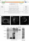
FIGURE 1.
FIGURE 1.
Characterization of antibodies against specific peptide sequences of human βIII spectrin. A, schematic representation of human βIII spectrin and targeted regions are indicated by arrows. Peptide 1 targets the ABD; peptide 2 targets the first spectrin repeat (Sp); peptides 3 and 4 target the nonspecific region; and peptide 5 targets the PH domain. The table shows the complete amino acid sequence of the five selected peptides. The names of obtained antibodies are also given. B, immunostaining of RPE1, COS-7, and NRK cells employing antibody 33. The images were analyzed by confocal microscopy. Bar, 10 μm. C, Western blotting analysis using preimmune serum or affinity purified antibody 82 of whole cell lysates from indicated cell types and Golgi-enriched fractions from rat liver. Arrowhead indicates the expected βIII spectrin band.
FIGURE 2.
FIGURE 2.
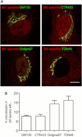
βIII spectrin is enriched in distal Golgi compartments. A, co-localization of βIII spectrin with Golgi markers. RPE1 cells were co-stained with anti-βIII spectrin antibody 33 and antibodies against cis- (GM130), medial- (CTR433), trans-Golgi (Golgin97), or the TGN (TGN46) proteins. Merge pixels are shown in white to facilitate the visualization of the co-labeling. Bar, 10 μm. B, quantitative analysis of the co-localization shown in A.
FIGURE 3.
FIGURE 3.
Depletion of βIII spectrin fragments the Golgi. (A). Silencing of βIII spectrin in RPE1 cells. Total cell lysate from RPE1 cells transfected for 96 h with control siRNA (non-targeting siRNA pool; lane 1) or βIII spectrin siRNA pool composed of four siRNAs (lane 2) or individualized siRNA (from 1 to 4; lanes 3–6, respectively) were subjected to immunoblot analysis using affinity-purified antibodies against βIII spectrin. Note the reduction of the lower electrophoretic band indicated with the arrowhead. Values are the mean ± S.E. from three independent experiments. Statistical significance according to one-way analysis of variance, using Bonferroni's multiple comparison test (***, p ≤ 0.001 in contrast to siRNA non-targeting). p190 RhoGAP was used as a loading control. B, fragmentation of the Golgi in βIII spectrin-depleted RPE1 cells. Cells transfected with control siRNA or βIII spectrin siRNA pool were stained with anti-βIII spectrin antibodies and the trans-Golgi marker Golgin97. Note the fragmented and variably dispersed Golgi phenotypes observed in βIII spectrin-depleted cells (asterisks). Bar, 10 μm. C, quantitative analysis of the fragmentation of the Golgi in control, βIII spectrin-depleted cells shown in B, and in knockdown cells expressing rat Myc-tagged βIII spectrin. The data show the mean ± S.E. from three independent experiments. Statistical significance according to one-way analysis of variance using Bonferroni's multiple comparison test; ***, p ≤ 0.001.
FIGURE 4.
FIGURE 4.
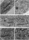
Ultrastructural alterations of the Golgi complex in βIII spectrin-silenced cells. RPE1 cells transfected with siRNA non-targeting (control) or βIII spectrin siRNA pools for 96 h were fixed and processed for conventional transmission electron microscopy. A, characteristic ultrastructure of the Golgi showing flat and attached cisternae in a control cell. B, panoramic area of the Golgi region in a knockdown cell showing Golgi stacks with variable ultrastructural alterations. One of these Golgi stacks is enlarged in C, in which distal cisternae are swollen. The arrowhead indicates the presence of a clathrin-coated vesicle. D and E, examples of Golgi fragments seen in silenced cells. Note the presence of distal swollen cisternae and/or tubulovesicular structures containing a large electrondense material (arrows). In some of these Golgi fragments, cisternae are not completely flat but remain attached (E). Box indicated in panel B is enlarged in panel C. Bars, 200 nm. G, Golgi complex: N, nucleus.
FIGURE 5.
FIGURE 5.
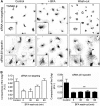
Depletion of βIII spectrin blocks the reassembly of the Golgi. Cells transfected with control or βIII spectrin siRNA pools were first treated with BFA (2.5 μg/ml for 90 min; +BFA). Thereafter, cells were washed out, fixed at different time points, and co-stained with antibodies to βIII spectrin (not shown) and to TGN46. Insets are representative images of enlarged cells. Bar, 10 μm. B, analysis of the Golgi compactness index in control (siRNA non-targeting) (left panel) and βIII spectrin knockdown cells (right panel) at different times after BFA wash-out. Values are the mean ± S.E. from three independent experiments (100 cells each). Statistical significance according to one-way analysis of variance using Bonferroni's multiple comparison test (*, p ≤ 0.05; ***, p ≤ 0.001). A.U., arbitrary units.
FIGURE 6.
FIGURE 6.
Depletion of βIII spectrin impairs the anterograde transport of the VSV-G and the post-Golgi secretion of 35S-labeled proteins. A, representative images of HeLa cells constitutively expressing VSV-G-GFP mutant form ts045 show the viral protein in the ER at 40 °C, or in the Golgi (30 min) or the plasma membrane (PM; 90 min) when cells were shifted to 32 °C. Bar, 10 μm. B, representative images of VSV-G-GFP expressing HeLa cells silenced with non-targeting or βIII spectrin siRNAs after 15 and 60 min after being shifted to 32 °C. C, quantitative analysis of results partially shown in B. The graph shows the percentage of the cells in which VSV-G-GFP is mainly visualized to the ER, to the Golgi, or to the plasma membrane. Data are shown as the mean ± S.E. of three independent experiments (100 cells each). D, biochemical transport assay for VSV-G-GFP using Endo H. HeLa cells constitutively expressing VSV-G-GFP were transfected for 96 h with siRNA control or against βIII spectrin and incubated at 40 °C for the last 24 h. Then, cells were shifted at 32 °C to induce the transport of VSV-G from the ER, lysed at indicated times, and subjected to Endo H treatment. R and S indicate Endo H-resistant and -sensitive forms, respectively. The ratio of the amount of Endo H-resistant form to that of total amount is plotted. Values are represented as the mean ± S.E. of three independent experiments. Statistical significance is shown, according to two-way analysis of variance using Bonferroni's post-tests (*, p ≤ 0.05). E, RPE1 cells were transfected with control or with βIII spectrin siRNA. Ninety-six h after the transfection, cells were pulse labeled with [35S]Met/Cys, incubated at 19 °C for 3 h and then shifted to 37 °C. At the indicated times, proteins in the culture supernatants and the cell lysates were precipitated and quantified by scintillation counting. As additional controls, the secretion assay was performed at 4 °C or in cells treated with BFA. Results are the mean ± S.E. from at least three independent experiments. Statistical significance according to two-way analysis of variance using Bonferroni's post-tests (**, p ≤ 0.01 and ***, p ≤ 0.001).
FIGURE 7.
FIGURE 7.
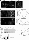
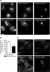
The association of βIII spectrin to Golgi membranes requires PI4P. A, COS-7 cells were transfected with the Golgi-targeted TGN38-FRB-CFP and the cytosolic mRFP12-FKBP only or with the cytosolic mRFP-FKBP12-Sac1 phosphatase. Live cells were preincubated for 5 min with rapamycin (100 nm), fixed, and incubated with antibodies against βIII spectrin. Bar, 10 μm. B, quantitative analysis of results shown in A. Results are the mean ± S.E. from three independent experiments using the Student's t test (***, p ≤ 0.001). C, RPE1 cells either expressing the GST-tagged PH domain of FAPP1 or the GFP-tagged PH domain of OSBP were fixed and stained with anti-GST and anti-βIII spectrin antibodies or only with anti-βIII spectrin antibodies, respectively. Note that cells with the Golgi-recruited PI4P Sac1 phophatase or with the presence of the PH domain of either FAPP1 or OSBP in the Golgi showed no staining for βIII spectrin (asterisks). Bar, 10 μm. A.U., arbitrary units.

Similar articles

Cited by

References

    1. Delaunay J. (2007) The molecular basis of hereditary red cell membrane disorders. Blood Rev. 21, 1–20 - PubMed
    1. Bennett V., Healy J. (2008) Organizing the fluid membrane bilayer: diseases linked to spectrin and ankyrin. Trends Mol. Med. 14, 28–36 - PubMed
    1. Machnicka B., Grochowalska R., Bogusławska D. M., Sikorski A. F., Lecomte M. C. (2012) Spectrin-based skeleton as an actor in cell signaling. Cell Mol. Life Sci. 69, 191–201 - PMC - PubMed
    1. Beck K. A., Buchanan J. A., Malhotra V., Nelson W. J. (1994) Golgi spectrin: identification of an erythroid β-spectrin homolog associated with the Golgi complex. J. Cell Biol. 127, 707–723 - PMC - PubMed
    1. Bennett V. (1989) The spectrin-actin junction of erythrocyte membrane skeletons. Biochim. Biophys. Acta 988, 107–121 - PubMed

Publication types

LinkOut - more resources