. 2013 Jan 1;190(1):270-7.
doi: 10.4049/jimmunol.1200061. Epub 2012 Dec 10.

IFN-γ from CD4 T cells is essential for host survival and enhances CD8 T cell function during Mycobacterium tuberculosis infection

Affiliations

IFN-γ from CD4 T cells is essential for host survival and enhances CD8 T cell function during Mycobacterium tuberculosis infection

Angela M Green et al. J Immunol. .

Abstract

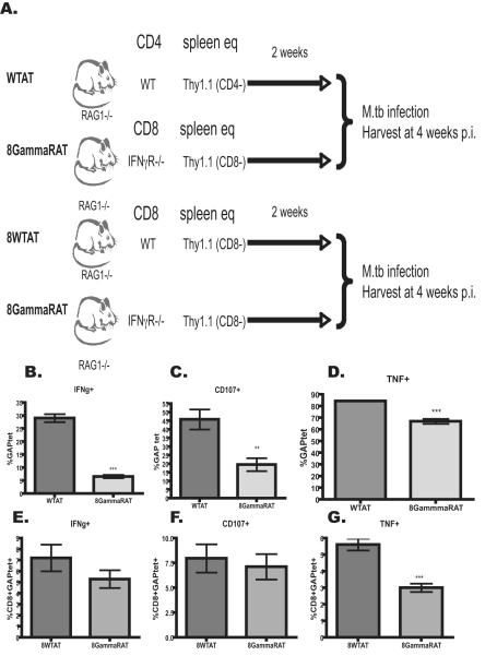
IFN-γ is necessary in both humans and mice for control of Mycobacterium tuberculosis. CD4 T cells are a significant source of IFN-γ during acute infection in mice and are required for control of bacterial growth and host survival. However, several other types of cells can and do produce IFN-γ during the course of the infection. We sought to determine whether IFN-γ from sources other than CD4 T cells was sufficient to control M. tuberculosis infection and whether CD4 T cells had a role in addition to IFN-γ production. To investigate the role of IFN-γ from CD4 T cells, a murine adoptive transfer model was developed in which all cells were capable of producing IFN-γ, with the exception of CD4 T cells. Our data in this system support that CD4 T cells are essential for control of infection, but also that IFN-γ from CD4 T cells is necessary for host survival and optimal long-term control of bacterial burden. In addition, IFN-γ from CD4 T cells was required for a robust CD8 T cell response. IFN-γ from T cells inhibited intracellular replication of M. tuberculosis in macrophages, suggesting IFN-γ may be necessary for intracellular bactericidal activity. Thus, although CD4 T cells play additional roles in the control of M. tuberculosis infection, IFN-γ is a major function by which these cells participate in resistance to tuberculosis.


Figures

Figure 1
Figure 1. Experimental design
A.) RAG1−/− mice were reconstituted with CD4-depleted splenocytes (Thy1.1) in the presence or absence (CTLAT) of CD4 T cells from either C57BL/6 (WTAT) or IFN-γ deficient (GammaAT) mice. B.) Prior to adoptive transfer, RAG1−/− mice were treated with probiotics to prevent development of inflammatory bowel disease (see Materials and Methods). Cells were adoptively transferred and mice allowed to reconstitute for 2.5 weeks. Mice were infected via aerosol wth low dose M.tuberculosis and harvested at serial time points. The experiments were repeated at least 3 times. C.) Representative flow cytometry plots of CD4 and CD8 T cells are shown for each experimental group (SSC vs CD4 or CD8) Representative plots for CD4 v IFN-γ and CD8 v IFN-γ following stimulation with anti-CD3/CD28 in the presence of monensin. D.) The frequency of CD4 and CD8 T cells within the live cell gate. E.) The frequency of macrophages (CD11b+CD11cdim), dendritic cell (CD11c+CD11b-) and neutrophils (GR-1+) within the live cell gate.
Figure 2
Figure 2. Granuloma in lungs of reconstituted mice
4 weeks p.i. one quarter of the lung was fixed, paraffin embedded and sectioned for H&E staining. Representative sections for each group are shown at 4X and 20X magnification.
Figure 3
Figure 3. CD4 T cells producing IFN-γ are needed for long-term survival of M. tuberculosis infection
A.) Adoptively transferred mice were followed for survival post-infection (N=5 per group), p=0.001, Log rank test. B.) Bacterial burden (CFU) in the lungs of adoptively transferred mice following M. tuberculosis infection; week 6: ANOVA p=0.002, Tukey’s multiple comparisons: **p<0.01 CTLAT v GammaAT; p<0.01 CTLAT v WTAT; Week 10: Student’s T test: GammaAT v WTAT **p=0.01. N=4/group/time point C.) Total number of cells in lungs at 4 weeks post infection; each symbol is one mouse from each group. ANOVA p=0.002; Tukey’s multiple comparisons **p<0.01 CTLAT v Gamma AT; **p<0.01 CTLAT v WTAT. Experiments were repeated at least three times.
Figure 4
Figure 4. Adoptively transferred CD4 T cells produce cytokines in the lungs in response to M. tuberculosis antigens
Cells from lung homogenates were stained with ESAT-6 tetramer and stimulated with ESAT-6 peptide (aa1-20) for 5 hours in monenesin, then for CD4 and Thy1.2.,and intracellular cytokines IL-2, IFN-γ, and TNF. A.) Representative plot: cells are gated on lymphocytes by size (forward and side scatter), then on Thy1.2 , then on CD4 and expressed as CD4 v ESAT-6 tetramer. B.) GammaAT and WTAT have similar frequencies of ESAT-6 tetramer+ cells in lungs at 4 weeks post-infection. C.) Only WTAT mice produce IFN-γ from CD4 T cells. GammaAT and WTAT CD4 T cells produce TNF (D) and IL-2 (E). As expected there are no CD4 T cells in the CTLAT lungs. N=4 mice/group. CD4+ESAT6tet+ T cells in both GammaAT and WTAT produced similar amounts of IL-2 (F) and TNF (G) per cell as measured by mean fluorescence intensity (MFI). Experiment was repeated at least 3 times.
Figure 5
Figure 5. CD8 T cell function is impaired in the absence of IFN-γ from CD4 T cells
Lung homogenates at 4 weeks post-infection were stimulated with GAPtetramer and GAP peptide and CD107 in the presence of monensin. Following stimulation cells were stained for CD8,Thy1.1., and intracellular cytokines TNF and IFN-γ. A.) Representative plots: live cell gate, lymphcyte gate, Thy1.1, CD8+ gate and GAPtet+ within the CD8+ gate. B.) WTAT lungs have more CD8+GAPtet+ cells compared to CTLAT lungs; ANOVA p=0.01, Tukey’s multiple comparisons: **p<0.01 CTLAT v WTAT. C. Number of CD8+ GAPtet+IFN-g+ T cells in lungs of WTAT, GammaAT and CTLAT mice. ANOVA p=0.015, Tukey’s *p<0.05 CTLAT v WTAT. D. Number of CD8+GAPtet+CD107+ cells in lungs; ANOVA p=0.008; Tukey’s *p<0.05 WTAT v CTLAT and GammaAT v WTAT. E. CD8+GAPtet+TNF+ cells in lungs; ANOVA p=0.015; Tukey’s *p<0.05 CTLAT v WTAT.
Figure 6
Figure 6. In the absence of IFN-γ the M. tuberculosis-specific CD8 T cell response is truncated
A.) Experimental design: RAG1−/− mice were reconstituted with CD8 depleted Thy1.1 splenocytes in the presence of CD8 T cells from or IFN-γR −/− mice and compared against WTAT (previously described in figure 1) at 4 weeks post infection, B) RAG1 −/− mice were received naïve CD8 T cells from either WT or IFN-γR−/− mice in combination with Thy 1.1 CD8 Depleted naïve splenocytes. CD8 T cells from experimental design shown in A were evaluated for C.) IFN-γ (students t-test ***p<0.0001), D.) CD107+ (student t-test **p<0.001) and E.) TNF (student t-test ***p<0.0001)production. CD8 T cells from experimental design shown in B were analyzed for F) IFNγ, G) CD107 and H) TNF. Experiments were completed at least 2 times.
Figure 7
Figure 7. IFN-γ signal is required to induce intracellular bacterial killing in macrophages
Infected bone marrow derived macrophages from C57BL/6 or IFNγR deficient mice were cultured for 72 hours with either media, IFN-Γ and LPS, CD4 T cells or CD8 T cells isolated from the lungs of M. tuberculosis infected mice. Data are reported as a percent killing ((CFU output/CFU input)×100)). (* p<0.05 Mann-whitney U test)

Similar articles

Cited by

References

    1. Lighvani AA, et al. T-bet is rapidly induced by interferon-gamma in lymphoid and myeloid cells. Proc Natl Acad Sci U S A. 2001;98(26):15137–42. - PMC - PubMed
    1. Saito S, Nakano M. Nitric oxide production by peritoneal macrophages of Mycobacterium bovis BCG-infected or non-infected mice: regulatory role of T lymphocytes and cytokines. J Leukoc Biol. 1996;59(6):908–15. - PubMed
    1. Casanova JL, Abel L. Genetic dissection of immunity to mycobacteria: the human model. Annu Rev Immunol. 2002;20:581–620. - PubMed
    1. Flynn JL, et al. An essential role for interferon gamma in resistance to Mycobacterium tuberculosis infection. J Exp Med. 1993;178(6):2249–54. - PMC - PubMed
    1. Scanga CA, et al. The inducible nitric oxide synthase locus confers protection against aerogenic challenge of both clinical and laboratory strains of Mycobacterium tuberculosis in mice. Infect Immun. 2001;69(12):7711–7. - PMC - PubMed

Publication types

MeSH terms

Substances