. 2012:18:2896-908.
Epub 2012 Dec 1.

Development of ex vivo organ culture models to mimic human corneal scarring

Affiliations

Development of ex vivo organ culture models to mimic human corneal scarring

Hélène Janin-Manificat et al. Mol Vis. 2012.

Abstract

Purpose: To develop ex vivo organ culture models of human corneal scarring suitable for pharmacological testing and the study of the molecular mechanisms leading to corneal haze after laser surgery or wounding.

Methods: Corneas from human donors were cultured ex vivo for 30 days, either at the air-liquid interface (AL) or immersed (IM) in the culture medium. Histological features and immunofluorescence for fibronectin, tenascin C, thrombospondin-1, and α-smooth muscle actin were graded from 0 to 3 for control corneas and for corneas wounded with an excimer laser. The effects of adding 10 ng/ml transforming growth factor-β1 (TGF-β1) to the culture medium and of prior complete removal of the epithelium and limbus, thus preventing reepithelialization, were also analyzed on wounded corneas. Collagen III expression was detected with real-time PCR.

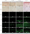
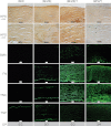
Results: Wounding alone was sufficient to induce keratocyte activation and stromal disorganization, but it was only in the presence of added TGF-β1 that intense staining for fibronectin and tenascin C was found in the AL and IM models (as well as thrombospondin-1 in the AL model) and that α-smooth muscle actin became detectable. The scar-like appearance of the corneas was exacerbated when TGF-β1 was added and reepithelialization was prevented, resulting in the majority of corneas becoming opaque and marked upregulation of collagen III.

Conclusions: THE MAIN FEATURES OF CORNEAL SCARRING WERE REPRODUCED IN THESE TWO COMPLEMENTARY MODELS: the AL model preserved differentiation of the epithelium and permits the topical application of active molecules, while the IM model ensures better perfusion by soluble compounds.


Figures

Figure 1
Figure 1
Conditions tested in this study. A: Details of procedures performed before subsequent culture, including removal of the epithelium and the outer part of the cornea including the limbus, leaving the epithelial basement membrane intact, and wounding using an excimer laser. B: Diagrammatic representation of the different culture conditions used, at the air-liquid interface (AL) and fully immersed in culture medium (IM). Conditions include non-wounded controls with the epithelium and limbus intact (represented by E) and wounded corneas (W) with and without prior removal of the epithelium and limbus, and with and without TGF-β1 (T) added at a concentration of 10 ng/ml.
Figure 2
Figure 2
Corneas cultured at the air-liquid interface. For each condition (E=control with the epithelium and the limbus intact; WE=wounded without prior removal of the epithelium and limbus; WET=as WE but with TGF-β1 added at 10 ng/ml; WT=wounded after prior removal of the epithelium and limbus plus TGF-β1 added at 10 ng/ml), keratocyte activation (see arrows) and stromal disorganization were assessed with hematoxylin phloxine saffron (HPS) staining (scale bars: 100 µm (row 1), 50 µm (row 2)), expression of α-smooth muscle actin (SMA), fibronectin (FN), tenascin C (TNC) and thrombospondin-1 (TSP) by immunofluorescence (scale bars=100 µm), and corneal opacity (OP) by the ability to read Arial 8 characters (number of opaque corneas/total number of corneas examined for each condition). Each figure shows the wound region only, except the control E, which shows the central part of the cornea.
Figure 3
Figure 3
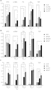
Semiquantitative evaluation of histochemical and immunofluorescence data. A: Corneas cultured at the air-liquid interface. The total numbers of corneas examined per group were: E=3, WE=3, WET=7; WT=6. B: Corneas cultured in immersed conditions. The total numbers of corneas examined per group were: E=3, WE=3, WET=3; WT=4. C: Comparison of semiquantitative data for control E and wounded corneas (WT; wounded after prior removal of the epithelium and limbus then cultured in the presence of added TGF-β1) between the air-liquid and immersed conditions. The total numbers of corneas examined per group were: AL/E=3, IM/E=3, AL/WT=6; IM/WT=4. In all cases, significant differences are indicated by * (p<0.05).
Figure 4
Figure 4
Assessment of corneal opacity. While corneas wounded without prior removal of the epithelium and the limbus (AL/WE, IM/WE) are transparent, corneas wounded after prior removal of the epithelium and limbus and cultured with added TGF-β1 (AL/WT, IM/WT) are opaque. Note that the latter corneas are smaller in diameter due to the total elimination of the limbus by trepanation (8 mm diameter trepan). Scale bars=5 mm.
Figure 5
Figure 5
Corneas cultured in fully immersed conditions. For each condition (E=control with the epithelium and limbus intact; WE=wounded without prior removal of the epithelium and limbus; WET=as WE but with TGF-β1 added at 10 ng/ml; WT=wounded after prior removal of the epithelium and limbus plus TGF-β1 added at 10 ng/ml), keratocyte activation (see arrows) and stromal disorganization were assessed with HPS staining (scale bars: 100 µm (row 1), 50 µm (row 2)), expression of α-smooth muscle actin (SMA), fibronectin (FN), tenascin C (TNC), and thrombospondin-1 (TSP) with immunofluorescence (scale bars=100 µm), and corneal opacity (OP) by the ability to read Arial 8 characters (number of opaque corneas / total number of corneas examined for each condition). Each figure shows the wound region only, except the control E, which shows the central part of the cornea.

Similar articles

Cited by

References

    1. Hayashida Y, Nishida K, Yamato M, Yang J, Sugiyama H, Watanabe K, Hori Y, Maeda N, Kikuchi A, Okano T, Tano Y. Transplantation of tissue-engineered epithelial cell sheets after excimer laser photoablation reduces postoperative corneal haze. Invest Ophthalmol Vis Sci. 2006;47:552–7. - PubMed
    1. Netto MV, Mohan RR, Sinha S, Sharma A, Gupta PC, Wilson SE. Effect of prophylactic and therapeutic mitomycin C on corneal apoptosis, cellular proliferation, haze, and long-term keratocyte density in rabbits. J Refract Surg. 2006;22:562–74. - PMC - PubMed
    1. Sharma A, Mehan MM, Sinha S, Cowden JW, Mohan RR. Trichostatin a inhibits corneal haze in vitro and in vivo. Invest Ophthalmol Vis Sci. 2009;50:2695–701. - PMC - PubMed
    1. Pellegrini G, Rama P, Mavilio F, De LM. Epithelial stem cells in corneal regeneration and epidermal gene therapy. J Pathol. 2009;217:217–28. - PubMed
    1. Liang L, Sheha H, Li J, Tseng SC. Limbal stem cell transplantation: new progresses and challenges. Eye (Lond) 2009;23:1946–53. - PMC - PubMed

Publication types

MeSH terms