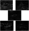
Ultrasonographic appearance of adrenal glands in healthy and sick cats
- PMID: 23234721
- PMCID: PMC10816315
- DOI: 10.1177/1098612X12469523
Ultrasonographic appearance of adrenal glands in healthy and sick cats
Abstract
The first part of the study aimed to describe prospectively the ultrasonographic features of the adrenal glands in 94 healthy cats and 51 chronically sick cats. It confirmed the feasibility of ultrasonography of adrenal glands in healthy and chronically sick cats, which were not statistically different. The typical hypoechoic appearance of the gland surrounded by hyperechoic fat made it recognisable. A sagittal plane of the gland, not in line with the aorta, may be necessary to obtain the largest adrenal measurements. The reference intervals of adrenal measurements were inferred from the values obtained in the healthy and chronically sick cats (mean ± 0.96 SD): adrenal length was 8.9-12.5 mm; cranial height was 3.0-4.8 mm; caudal height was 3.0-4.5 mm. The second part of the study consisted of a retrospective analysis of the ultrasonographic examination of the adrenal glands in cats with adrenal diseases (six had hyperaldosteronism and four had pituitary-dependent hyperadrenocorticism) and a descriptive comparison with the reference features obtained in the control groups from the prospective study. Cats with hyperaldosteronism presented with unilateral severely enlarged adrenal glands. However, a normal contralateral gland did not preclude a contralateral infiltration in benign or malignant adrenal neoplasms. The ultrasonographic appearance of the adrenal glands could not differentiate benign and malignant lesions. The ultrasonographic appearance of pituitary-dependent hyperadrenocorticism was mainly a symmetrical adrenal enlargement; however, a substantial number of cases were within the reference intervals of adrenal size.
Conflict of interest statement
The authors do not have any potential conflicts of interest to declare.
Figures


References
-
- Cartee RE, Finn Bodner ST, Gray BW. Ultrasound examination of the feline adrenal gland. J Diagn Med Ultrasound 1993; 9: 327–330.
-
- Zimmer C, Hörauf A, Reusch C. Ultrasonographic examination of the adrenal gland and evaluation of the hypophyseal-adrenal axis in 20 cats. J Small Anim Pract 2000; 41:156–160. - PubMed
-
- Zatelli A, D’Ippolito P, Fiore I, Zini E. Ultrasonographic evaluation of the size of the adrenal glands of 24 diseased cats without endocrinopathies. Vet Rec 2007; 160: 658–660. - PubMed
-
- Westropp JI, Welk KA, Tony Buffington CA. Small adrenal glands in cats with feline interstitial cystitis. J Urol 2003; 170: 2494–2497. - PubMed
-
- Kley S, Alt M, Zimmer C, Hoerauf A, Reusch CE. Evaluation of the low-dose dexamethasone suppression test and ultrasonographic measurements of the adrenal glands in cats with diabetes mellitus. Schweiz Arch Tierheilkd 2007; 149: 493–500. - PubMed
MeSH terms
LinkOut - more resources
Full Text Sources
Medical
Miscellaneous

