

Epigenetic regulation of planarian stem cells by the SET1/MLL family of histone methyltransferases
- PMID: 23235145
- PMCID: PMC3549883
- DOI: 10.4161/epi.23211
Epigenetic regulation of planarian stem cells by the SET1/MLL family of histone methyltransferases
Abstract
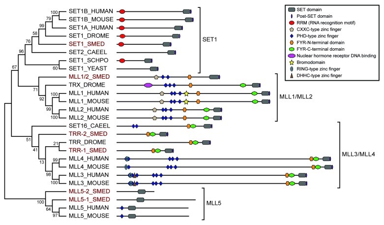
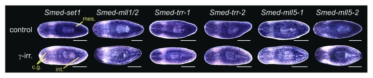
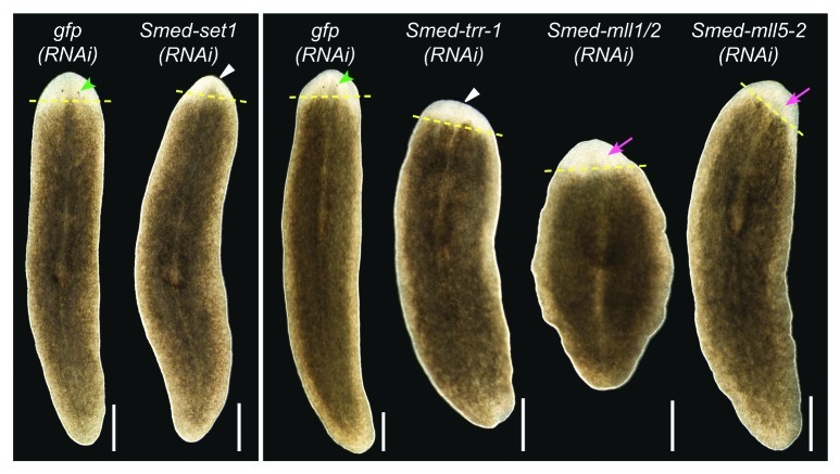
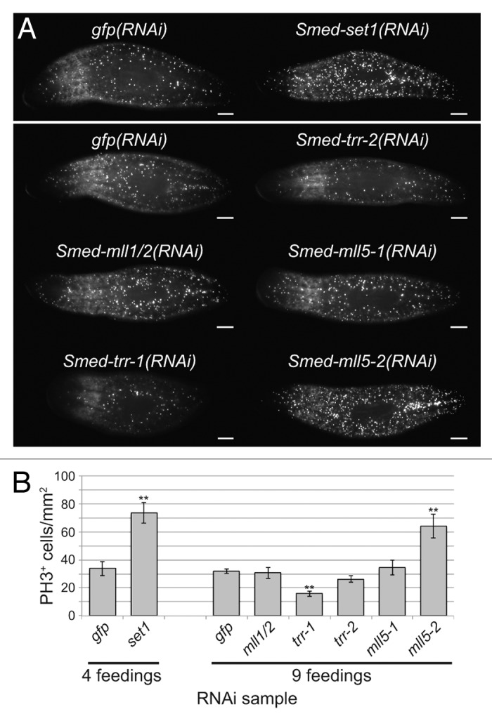
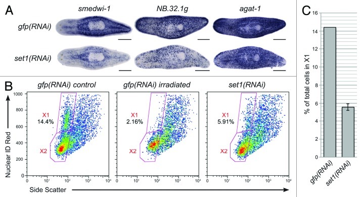
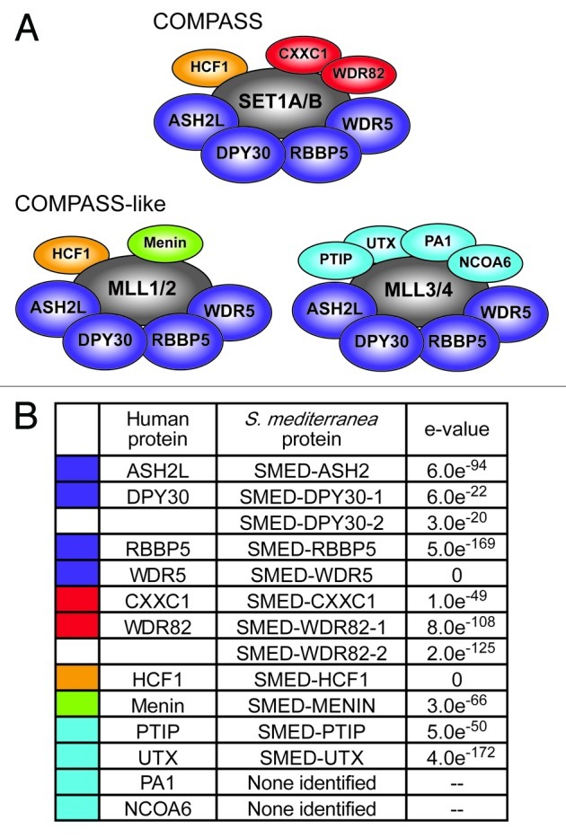
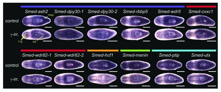
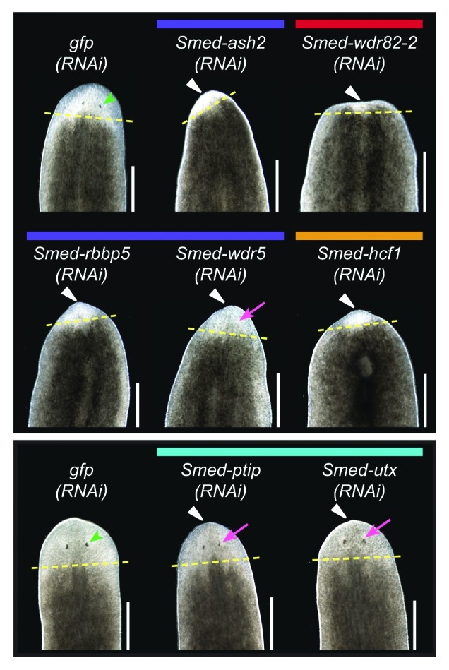
Chromatin regulation is a fundamental mechanism underlying stem cell pluripotency, differentiation, and the establishment of cell type-specific gene expression profiles. To examine the role of chromatin regulation in stem cells in vivo, we study regeneration in the freshwater planarian Schmidtea mediterranea. These animals possess a high concentration of pluripotent stem cells, which are capable of restoring any damaged or lost tissues after injury or amputation. Here, we identify the S. mediterranea homologs of the SET1/MLL family of histone methyltransferases and COMPASS and COMPASS-like complex proteins and investigate their role in stem cell function during regeneration. We identified six S. mediterranea homologs of the SET1/MLL family (set1, mll1/2, trr-1, trr-2, mll5-1 and mll5-2), characterized their patterns of expression in the animal, and examined their function by RNAi. All members of this family are expressed in the stem cell population and differentiated tissues. We show that set1, mll1/2, trr-1, and mll5-2 are required for regeneration and that set1, trr-1 and mll5-2 play roles in the regulation of mitosis. Most notably, knockdown of the planarian set1 homolog leads to stem cell depletion. A subset of planarian homologs of COMPASS and COMPASS-like complex proteins are also expressed in stem cells and implicated in regeneration, but the knockdown phenotypes suggest that some complex members also function in other aspects of planarian biology. This work characterizes the function of the SET1/MLL family in the context of planarian regeneration and provides insight into the role of these enzymes in adult stem cell regulation in vivo.
Keywords: COMPASS; H3K4; MLL; SET1; histone methyltransferase; neoblasts; planarian; regeneration; stem cells.
Figures
Similar articles
-
CBP/p300 homologs CBP2 and CBP3 play distinct roles in planarian stem cell function.Dev Biol. 2021 May;473:130-143. doi: 10.1016/j.ydbio.2021.02.004. Epub 2021 Feb 16. Dev Biol. 2021. PMID: 33607113
-
Chromatin remodeling protein BPTF mediates chromatin accessibility at gene promoters in planarian stem cells.BMC Genomics. 2025 Mar 11;26(1):232. doi: 10.1186/s12864-025-11405-3. BMC Genomics. 2025. PMID: 40069606 Free PMC article.
-
Planarians as a model to assess in vivo the role of matrix metalloproteinase genes during homeostasis and regeneration.PLoS One. 2013;8(2):e55649. doi: 10.1371/journal.pone.0055649. Epub 2013 Feb 6. PLoS One. 2013. PMID: 23405188 Free PMC article.
-
Decoding Stem Cells: An Overview on Planarian Stem Cell Heterogeneity and Lineage Progression.Biomolecules. 2021 Oct 17;11(10):1532. doi: 10.3390/biom11101532. Biomolecules. 2021. PMID: 34680165 Free PMC article. Review.
-
Planarian flatworms as a new model system for understanding the epigenetic regulation of stem cell pluripotency and differentiation.Semin Cell Dev Biol. 2019 Mar;87:79-94. doi: 10.1016/j.semcdb.2018.04.007. Epub 2018 Apr 27. Semin Cell Dev Biol. 2019. PMID: 29694837 Review.
Cited by
-
m6 A promotes planarian regeneration.Cell Prolif. 2023 May;56(5):e13481. doi: 10.1111/cpr.13481. Epub 2023 Apr 21. Cell Prolif. 2023. PMID: 37084418 Free PMC article.
-
A functional genomics screen identifies an Importin-α homolog as a regulator of stem cell function and tissue patterning during planarian regeneration.BMC Genomics. 2015 Oct 12;16:769. doi: 10.1186/s12864-015-1979-1. BMC Genomics. 2015. PMID: 26459857 Free PMC article.
-
Conservation of epigenetic regulation by the MLL3/4 tumour suppressor in planarian pluripotent stem cells.Nat Commun. 2018 Sep 7;9(1):3633. doi: 10.1038/s41467-018-06092-6. Nat Commun. 2018. PMID: 30194301 Free PMC article.
-
Biochemical reconstitution and phylogenetic comparison of human SET1 family core complexes involved in histone methylation.J Biol Chem. 2015 Mar 6;290(10):6361-75. doi: 10.1074/jbc.M114.627646. Epub 2015 Jan 5. J Biol Chem. 2015. PMID: 25561738 Free PMC article.
-
Interaction of DNA demethylase and histone methyltransferase upregulates Nrf2 in 5-fluorouracil-resistant colon cancer cells.Oncotarget. 2016 Jun 28;7(26):40594-40620. doi: 10.18632/oncotarget.9745. Oncotarget. 2016. PMID: 27259240 Free PMC article.
References
Publication types
MeSH terms
Substances
Grants and funding
LinkOut - more resources
Full Text Sources
Other Literature Sources
Medical