. 2013 Feb 1;288(5):3641-54.
doi: 10.1074/jbc.M112.433615. Epub 2012 Dec 12.

Accelerated disease onset with stabilized familial amyotrophic lateral sclerosis (ALS)-linked mutant TDP-43 proteins

Affiliations

Accelerated disease onset with stabilized familial amyotrophic lateral sclerosis (ALS)-linked mutant TDP-43 proteins

Shoji Watanabe et al. J Biol Chem. .

Abstract

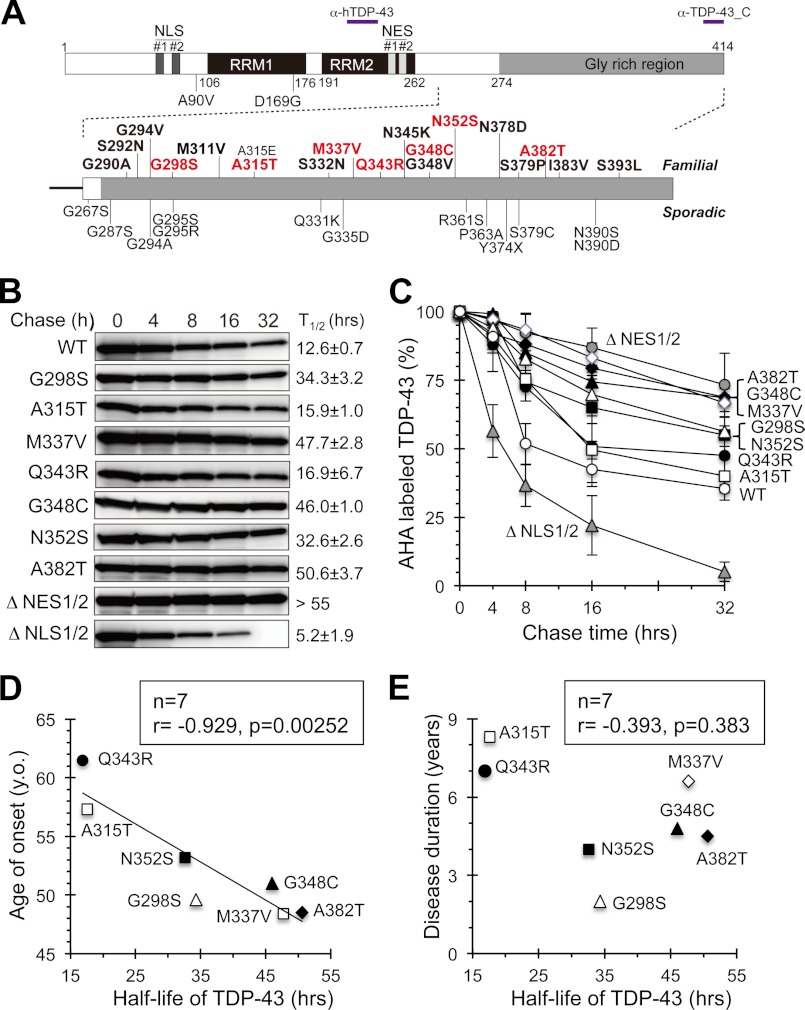
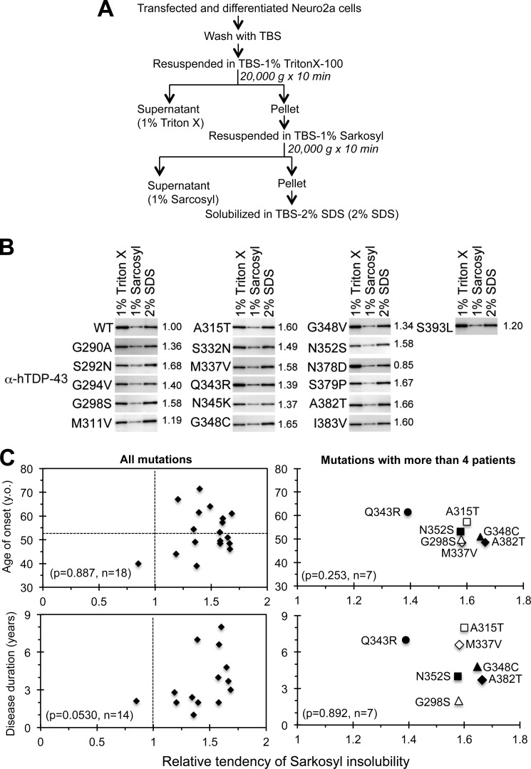
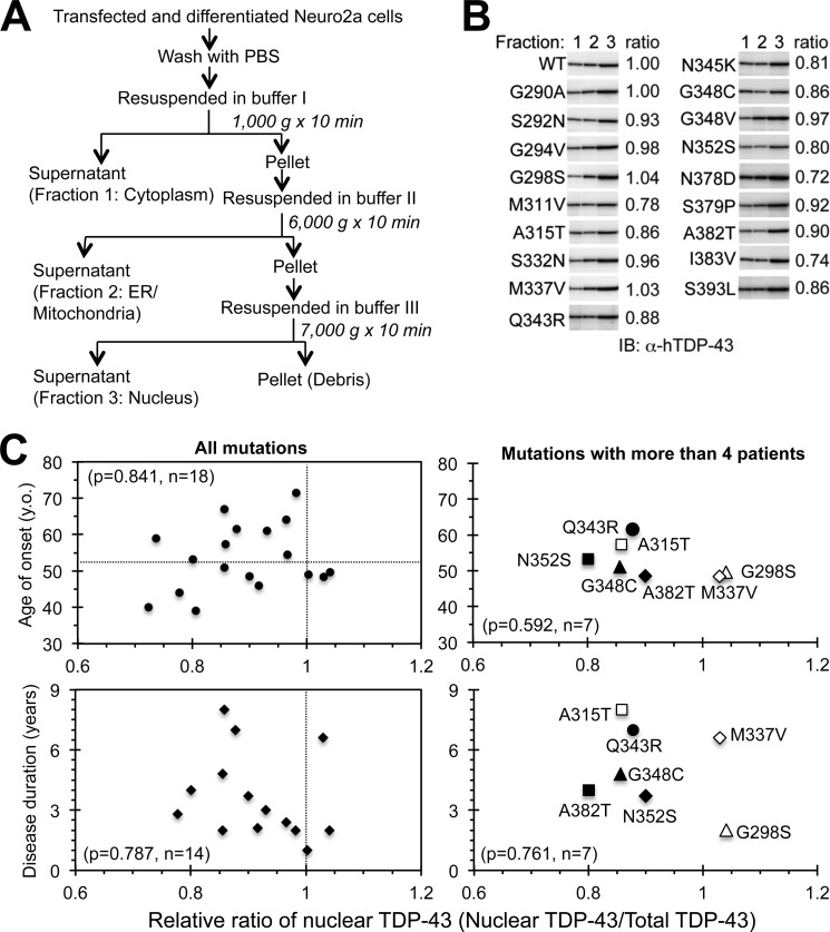
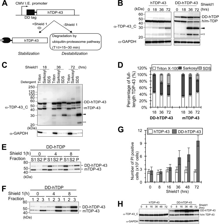
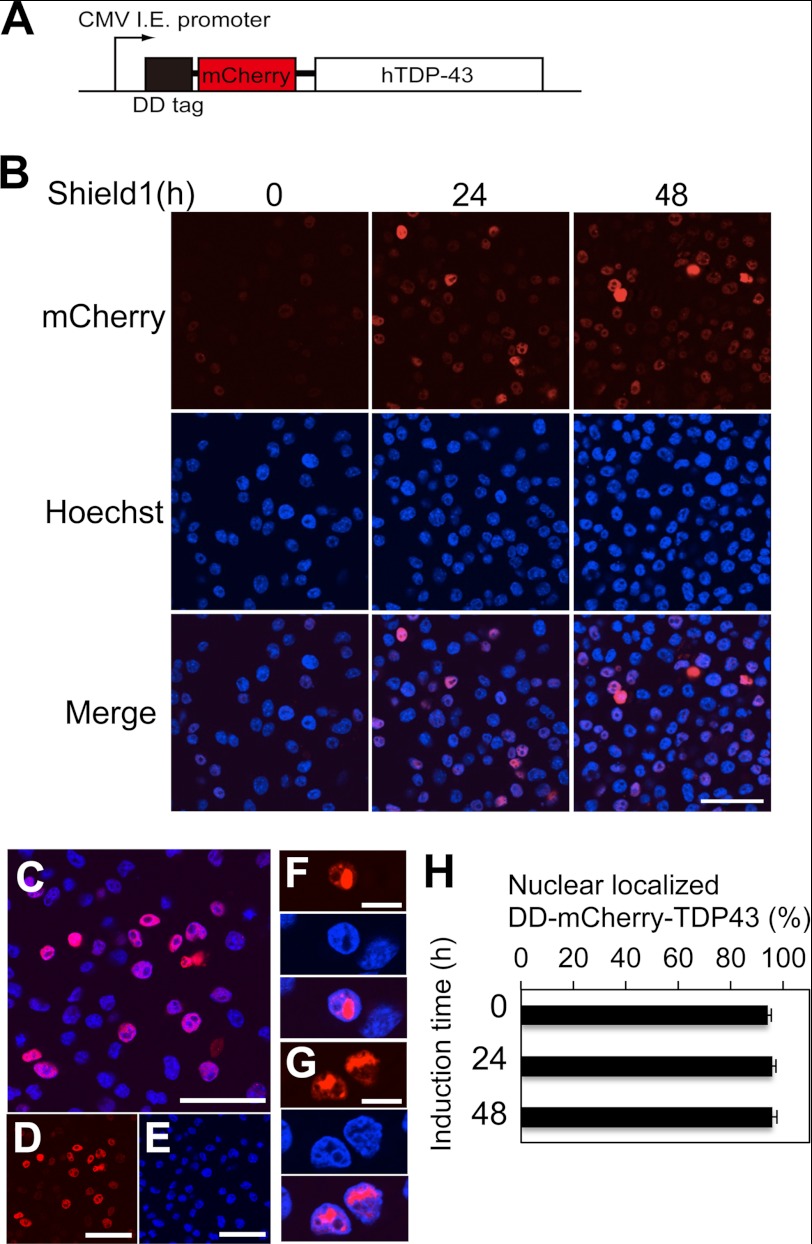
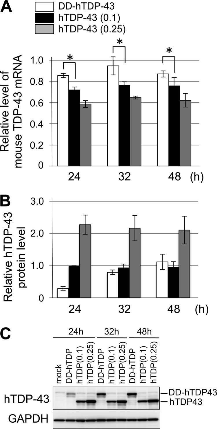
Abnormal protein accumulation is a pathological hallmark of neurodegenerative diseases, including accumulation of TAR DNA-binding protein 43 (TDP-43) in amyotrophic lateral sclerosis (ALS). Dominant mutations in the TDP-43 gene are causative for familial ALS; however, the relationship between mutant protein biochemical phenotypes and disease course and their significance to disease pathomechanism are not known. Here, we found that longer half-lives of mutant proteins correlated with accelerated disease onset. Based on our findings, we established a cell model in which chronic stabilization of wild-type TDP-43 protein provoked cytotoxicity and recapitulated pathogenic protein cleavage and insolubility to the detergent Sarkosyl, TDP-43 properties that have been observed in sporadic ALS lesions. Furthermore, these cells showed proteasomal impairment and dysregulation of their own mRNA levels. These results suggest that chronically increased stability of mutant or wild-type TDP-43 proteins results in a gain of toxicity through abnormal proteostasis.


Figures

FIGURE 1.
FIGURE 1.
Early disease onset correlates with increased stability of mutant TDP-43 proteins in familial ALS. A, schematic drawing of human TDP-43 and its mutations. ALS-linked mutations of TDP-43 from familial (upper) and sporadic (lower) ALS cases as well as the locations of the NLS and NES are shown. The mutations used in our study are shown in boldface type (almost all familial ALS-linked mutations), and mutations with more than four patients are shown in red. The epitopes recognized by the anti-TDP-43 antibodies used in this study are indicated. B and C, half-lives of wild-type and mutant TDP-43 proteins. B, transfected Neuro2a cells were metabolically labeled with the methionine analog AHA. Labeled TDP-43 was immunoprecipitated and visualized by HRP-streptavidin as described under “Experimental Procedures.” C, labeled TDP-43 bands were quantified, and the averages of three independent experiments were plotted. Half-lives of wild-type and mutant TDP-43 proteins were calculated by curve-fitting using this graph (B, right). Error bars represent S.E. D and E, half-lives of familial ALS-linked mutant TDP-43 proteins were negatively correlated with age of disease onset but not disease duration. Calculated half-lives of TDP-43 proteins were plotted against mean ages of disease onset (D) or duration (E). The correlations between each parameter and age of onset or duration were evaluated by the correlation coefficient (r) and probability (p) shown in each graph (D and E).
FIGURE 2.
FIGURE 2.
Detergent insolubility of ALS-linked TDP-43 mutants is not correlated with onset or duration of disease. A and B, determination of Sarkosyl solubility of exogenous human TDP-43 in differentiated Neuro2a cells. Neuro2a cells were transiently transfected with wild-type or ALS-linked mutant TDP-43 expression vectors and differentiated for 24 h. The solubility of human TDP-43 was determined by differential solubilization using three different detergents (Triton X-100, Sarkosyl, and SDS) as illustrated (A). Each fraction was analyzed by immunoblotting with anti-human TDP-43 antibody (B). C, solubility of ALS-linked TDP-43 mutants did not correlate with onset or duration of disease. The ratio of the amount of TDP-43 in the Sarkosyl-insoluble fraction (SDS-soluble fraction) to the total amount was normalized using the ratio of wild-type TDP-43 (B, right). Mean values from three independent experiments were used to calculate a relative tendency of Sarkosyl insolubility. Mean onset (C, upper panels) and duration (C, lower panels) of all familial ALS-linked mutations (C, left panels) or mutations in a significant number of patients (more than four) (C, right panels) were plotted against the relative ratios of Sarkosyl insolubility. The significance of correlation was evaluated by the probability (p value) shown in each graph. y.o., years old.
FIGURE 3.
FIGURE 3.
Subcellular localization of TDP-43 is not correlated with disease onset or duration of mutant TDP-43-mediated ALS. A and B, subcellular fractionation of differentiated Neuro2a cells expressed human TDP-43. Neuro2a cells were transiently transfected with wild-type or ALS-linked mutant TDP-43 expression vectors and differentiated with Bt2cAMP. Cells were fractionated by differential centrifugation as illustrated (A). Subsequently, proteins in each fraction were analyzed by immunoblotting (IB) using anti-human TDP-43 antibody (B). C, subcellular localization of mutant TDP-43 proteins had no correlation with the time of disease onset or the disease duration. The ratio of nuclear fraction per total amount of TDP-43 protein in each mutant was calculated, normalized with the ratio of wild-type protein (B), and plotted with the mean onset age (C, upper panel) and the mean duration (C, lower panel) of each mutation. All familial ALS-linked TDP-43 mutations (C, left panel), or mutations with a significant number of patients (more than four) (C, right panel) were plotted. The significance of correlation was evaluated by the probability (p value) shown in each graph. y.o., years old.
FIGURE 4.
FIGURE 4.
Increased stability of TDP-43 leads to protein cleavage, detergent insolubility, and cytotoxicity in neuronal cells. A, schematic diagram of the expression vector to control TDP-43 stability. I.E., immediate early enhancer. B, cleavage of TDP-43 protein was increased by stabilized DD-fused TDP-43. Neuro2a cells were transfected with nontagged (hTDP-43) or DD-fused TDP-43 (DD-TDP-43), differentiated, and then incubated with Shield1 for the indicated time. Total protein extracts were analyzed by immunoblotting using antibodies directed against the TDP-43 C terminus (TDP-43_C) and GAPDH. Cleaved fragments of TDP-43 migrating to ∼30 and 25 kDa are indicated with black and white arrowheads, respectively. C and D, detergent solubility of TDP-43 was decreased by stabilization of proteins. After stabilization for the indicated time, cells were sequentially solubilized by the indicated detergents. Each fraction was analyzed by immunoblotting using anti-TDP-43_C and GAPDH antibodies (C). D, percentage of full-length DD-TDP-43 or endogenous TDP-43 in each fraction measured in C was quantified. E and F, DD-hTDP-43 protein shares the similar properties with the endogenous wild-type TDP-43 during short term accumulation. Neuro2a cells were transiently transfected with DD-hTDP-43. DD-hTDP-43 was stabilized by incubation with Shield 1 for the indicated time. E, solubility of TDP-43 to the detergents was examined by the same method shown in Fig. 2. S1, 1% Triton X-100-soluble fraction; S2, 1% Sarkosyl-soluble fraction; P, 2% SDS-soluble fraction. F, subcellular localization of TDP-43 was analyzed by the same method shown in Fig. 3. Fraction 1, 2, and 3 indicate cytoplasm, endoplasmic reticulum/mitochondria, and nuclear fraction, respectively. E and F, each fraction was analyzed by immunoblotting using antibody against the TDP-43 C terminus (TDP-43_C). G and H, cytotoxicity was induced by stabilized TDP-43. Nontagged TDP-43 or DD-TDP-43 was transfected into Neuro2a cells. After incubation with Shield1 for the indicated time, cells were stained with PI. PI-positive cells were counted by FACS analysis (G). Expression levels of TDP-43 under the same conditions as G were confirmed by immunoblotting (H). Averages from three (D) or six (G) independent experiments are plotted, and error bars represent S.E.
FIGURE 5.
FIGURE 5.
Stabilized full-length TDP-43 proteins predominantly localize to the nucleus. A, scheme for the plasmid encoding TDP-43 fused with both DD and mCherry (DD-mCherry-TDP-43) is illustrated. B–H, DD-mCherry-TDP-43 was stabilized by incubation with Shield1 for the indicated time in differentiated Neuro2a cells. DD-mCherry-TDP-43 was visualized by confocal microscopy. I.E., immediate early enhancer. B–G, representative image of cells expressing stabilized DD-mCherry-TDP-43. B, representative confocal images of cells with stabilized TDP-43 for the indicated time visualized with mCherry, Hoechst, or both. C–E, confocal images of cells with stabilized TDP-43 for 24 h visualized with mCherry and Hoechst (C), mCherry (D), or Hoechst (E). F and G, cells with nuclear aggregates containing stabilized DD-mCherry-TDP-43 were visualized with mCherry (top panel), Hoechst (middle panel), or both (bottom panel). H, percentage of cells expressing nuclear TDP-43 among total mCherry-TDP-43 positive cells was calculated. Averages from three independent experiments are plotted. Bars represent S.E. Bars B–E, 50 μm; F and G, 10 μm.
FIGURE 6.
FIGURE 6.
Dysregulation of TDP-43 mRNA is caused by increased stability of wild-type TDP-43 protein. A, reverse transcription-PCR analysis of endogenous mouse TDP-43 mRNA in differentiated Neuro2a cells in the presence of DD-TDP-43 (white bar), hTDP-43 with transfection of 0.1 μg of plasmid (0.1; black bar), or hTDP-43 with transfection of 0.25 μg of plasmid (0.25; gray bar). X axis indicates the time after the initial transfection of the plasmids. Y axis denotes the levels of mouse TDP-43 mRNA relative to those in mock-transfected cells. The mean value from four independent experiments was plotted.*, p < 0.01 (Student's t test). B and C, expression levels of exogenous DD-hTDP-43 and hTDP-43 proteins in the cells in A were measured by immunoblots using anti-hTDP-43 antibody (C). The quantification of DD-hTDP-43 and hTDP-43 proteins normalized to the levels of GAPDH was plotted (B).
FIGURE 7.
FIGURE 7.
Exon skipping activity is preserved in familial ALS-linked mutant TDP-43. A, schematic drawing of CFTR mini-gene construct, which was developed with a modification of pTG11T5-hCFTR exon9, was illustrated (A, left). The size of PCR product after reverse transcription including (+) or excluding (−) exon9 is ∼420 or 240 bp, respectively (A, right). B, Neuro2a cells were transiently co-transfected with both hTDP-43 and hCFTR mini-gene expression plasmids and then differentiated with Bt2cAMP. Total RNAs were extracted from transfected cells, and cDNAs of hCFTR mini-gene including or excluding exon9 were amplified by the indicated primer pair (a2, Bra) after reverse transcription. The amplified PCR products were separated by electrophoresis using 1.5% agarose gel (B, top). To confirm expression levels of hTDP-43, total protein extracts were analyzed by immunoblotting using antibodies for human TDP-43 (B, middle) and GAPDH (B, bottom). M.M., molecular marker. C, amounts of hCFTR PCR products, excluding exon9 in B, compared with wild-type TDP-43-transfected cells were plotted. Mean values from three independent experiments were plotted, and error bars represent S.E.
FIGURE 8.
FIGURE 8.
Stabilized TDP-43 induces impairment of proteasome activity. A, schematic drawing of the vector (UbG76V-AcGFP) for monitoring cellular proteasome activity. To facilitate proteasomal degradation of UbG76V-AcGFP in cells, four lysine residues were introduced to the linker sequence between mutant ubiquitin and AcGFP. B and C, proteasome activity was impaired by stabilized TDP-43. B, Neuro2a cells were co-transfected with DD-TDP-43 and UbG76V-AcGFP plasmids. DD-TDP-43 protein was stabilized using the same method shown in Fig. 3. Total cell extract was analyzed by immunoblotting using antibodies directed against AcGFP, TDP-43 C-terminal (TDP-43_C), and GAPDH. As a control experiment, Neuro2a cells were transfected with UbG76V-AcGFP in the absence or presence of 0.2 μm MG132 for 72 h (right). C, expression levels of UbG76V-AcGFP relative to that at 0 h are plotted. D, Neuro2a cells were co-transfected with UbG76V-AcGFP and hTDP-43 or DD-hTDP-43 using the same method shown in B. The levels of AcGFP, TDP-43, and GAPDH were analyzed by immunoblots. E, cleaved TDP-43 accumulated by proteasome inhibition, not autophagic inhibition. DD-TDP-43 was stabilized in Neuro2a cells, and the cells were then treated with the indicated reagents (UT, 0.1% DMSO; MG, 1 μm MG132; Epo, 0.25 μm epoxomicin; Wor, 0.1 μm wortmannin; Rap, 0.5 μm rapamycin; Baf, 0.1 μm bafilomycin A1) for 16 h. After treatment, total protein extracts were analyzed by immunoblotting using antibodies directed against TDP-43_C, p62/SQSTM1, LC3, and GAPDH (E). TDP-43 fragments of 30 and 25 kDa are indicated with black and white arrowheads, respectively.

Similar articles

Cited by

References

    1. Neumann M., Sampathu D. M., Kwong L. K., Truax A. C., Micsenyi M. C., Chou T. T., Bruce J., Schuck T., Grossman M., Clark C. M., McCluskey L. F., Miller B. L., Masliah E., Mackenzie I. R., Feldman H., Feiden W., Kretzschmar H. A., Trojanowski J. Q., Lee V. M. (2006) Ubiquitinated TDP-43 in frontotemporal lobar degeneration and amyotrophic lateral sclerosis. Science 314, 130–133 - PubMed
    1. Arai T., Hasegawa M., Akiyama H., Ikeda K., Nonaka T., Mori H., Mann D., Tsuchiya K., Yoshida M., Hashizume Y., Oda T. (2006) TDP-43 is a component of ubiquitin-positive tau-negative inclusions in frontotemporal lobar degeneration and amyotrophic lateral sclerosis. Biochem. Biophys. Res. Commun. 351, 602–611 - PubMed
    1. Chen-Plotkin A. S., Lee V. M., Trojanowski J. Q. (2010) TAR DNA-binding protein 43 in neurodegenerative disease. Nat. Rev. Neurol. 6, 211–220 - PMC - PubMed
    1. Cohen T. J., Lee V. M., Trojanowski J. Q. (2011) TDP-43 functions and pathogenic mechanisms implicated in TDP-43 proteinopathies. Trends Mol. Med. 17, 659–667 - PMC - PubMed
    1. Lagier-Tourenne C., Cleveland D. W. (2009) Rethinking ALS. The FUS about TDP-43. Cell 136, 1001–1004 - PMC - PubMed

Publication types

MeSH terms